The Mustang Hoof
Mustangs are well known for their tough, durable hooves. Based on visual examination and X-ray radiographs, defects are uncommon. In his study of approximately 1800 mustangs from the Nevadan desert, Dr. Ric Redden, NANRIC INC., notes the complete absence of club feet, which are commonly observed in domestic light breeds (www.nanric.com/thewildhorsesfoot.asp). The most serious hoof defects in mustangs are probably eliminated by natural selection if the horse remains in the wild for a number of years. Note that there is not one unique type of mustang foot. The foot is very adaptable and its anatomy will vary depending on environment. For example, the feet of a mustang living in the rocky dry desert will differ from those in a more humid climate with less abrasive terrain. Based on this reasoning, domesticated mustangs that graze pastures in the eastern U.S. should quickly loose the distinctive hoof characteristics and conformation developed over years in the western high plains.
Recent research on the Australian Brumby, the counterpart of the American Mustang in the Southern Hemisphere, provides a good scientific basis for the effect of environment on hoof conformation (www.wildhorseresearch.com). This work also includes results from the microscopic examination of the internal hoof anatomy (histopathology), which indicates less than optimal foot health for feral horses living on hard, rocky terrains. For more information on studies of the Australian Bumbry refer to Chapter 2, The Feral Horse Foot - The Australian Brumby Studies, Brian Hampson, Ph.D., in the new book by Pete Ramey, Care and Rehabilitation of the Equine Foot, 2011.
Environment is obviously important in shaping the foot but genetics must also play a role. Some of the most convincing evidence for a genetic adaptation in the feral horse hoof comes from a comparative study of the fetuses of the Australian Brumby and domestic Thoroughbreds. Hampson et al. (2011) dissected the hoofs of near term fetuses from brumbies culled from herds in Northern Queensland (Lawn Hill) and Central Australia (Kings Canyon). The horses studied traveled on hard rocky ground and had been isolated with minimal intervention for about 100 years. The primary epidermal laminar density (PELD) in sections around the perimeter of the hoof wall of the feral fetuses show significant differences, with the density decreasing from the toe to the heel of the hoof. This is the same density variation observed in the hooves of adult horses. In contrast, PELD sections in the fetuses of domestic Thoroughbreds were uniform in density. The authors suggest that the distribution of the laminar density in the hooves of feral fetuses is a genetic preadaptation, which enables young foals to travel long distances in extreme environments soon after birth.
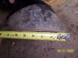
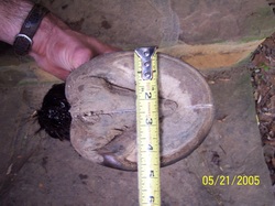
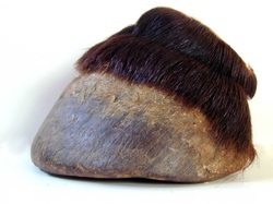
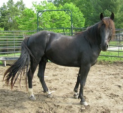
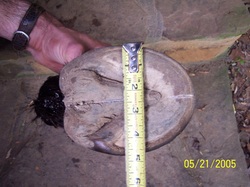
I was interested in comparing the feet of my domesticated mustangs with those of a wild mustang of approximately the same size and from the same general region. My horses have been removed from the western high plains for several years and, until recently, have been trimmed using conventional barefoot methods (pasture trim). I discovered some work by Pete Ramey (www.hoofrehab.com), who studied a cadaver foot from a well-preserved carcass of a filly found in Nevada after the spring thaw. The foot was frozen two days after collection, removed briefly for X-ray radiography and then refrozen before dissection. A lateral view of the front left hoof and the underside of the hoof are shown below (images courtesy of Pete Ramey).
The short hoof capsule and the "mustang roll", the rounded edge or bevel on the hoof wall, are characteristic of hooves shaped by the rocky, hilly terrain of the Nevadan desert. Note the concave shape of the sole, and the poor distinction between the frog and sole. Both the central sulcus and lateral sulci are shallow and not as deep as in domestic breeds. The grinding action of the terrain has literally polished the "mustang roll" and produced a very smooth, hard surface on the underside of the hoof. In some Nevada herds, the soles are flat with calloused heels, indicating that these mustangs walk on the bulbs of their heels (Dr. Ric Redden, pers. comm.).
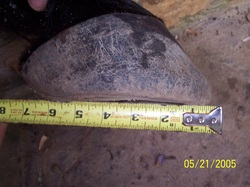
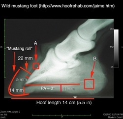
An image of the left front cadaver foot is compared with its digital radiograph below. The hoof length measured above (5.5 in, 14 cm) was used to calibrate the radiograph, which was annotated and measured using OsiriX for Mac OS X (GA Lager). The square labeled "B" denotes the lateral sulcus. The bright curved line represents a wire inserted into the sulcus to more precisely determined its position relative to the ground surface. The square labeled "A" defines the zone of dermal laminae, which are attached to the dorsal surface of the coffin bone and supply blood to the hoof. The dermal laminae interlock with the outer, epithermal laminae, creating a strong bond between the coffin bone and the hoof wall.
Comparison of LF cadaver foot with its digital X-ray radiograph (University of Georgia). Images courtesy of Pete Ramey. See text for explanation of symbols. Image processing, annotations and measurements by GA Lager, OsiriX for Mac OS X. Calibration of radiograph based on hoof length (5.5 in/14 cm), as measured by tape measure in the above image.
The radiograph illustrates some of the unique characteristics of the internal anatomy of a mustang hoof: 1. ground-parallel coffin bone (palmar angle (PA) ~ 0° on loading) 2. short hoof capsule with good sole depth (11-14 mm); 3. wide horn-laminar zone (HL) of uniform thickness (22 mm) 4. thick hoof wall. The wider HL zone in mature mustangs relative to light domestic breeds (15/15 mm) is due to the thicker hoof wall, which is about 2X the width of the dermal laminar zone (7.5 mm) (ref. Dr. Ric Redden). The HL zone in these horses usually reverts back to 15/15 mm several years after domestication. The somewhat smaller thickness of the laminae zone (5 mm) in the cadaver foot might be attributed to drying and contraction of the foot prior to X-ray radiography. This applies in particular to the laminae closest to the coffin bone, which on dissection were hardened and no longer elastic (Pete Ramey, pers. comm.).
|
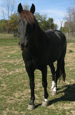
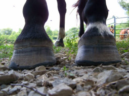
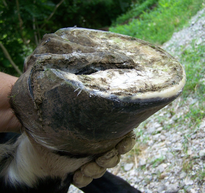
Good views of the hooves of Mesteño, my 5-year old gelding. He has beautiful hooves, very large, round and hard. In the pasture trim, the hoof wall bears most of the weight. The heel is trimmed back to the widest part of the frog, which is in contact with the ground surface when the hoof is loaded. The hoof wall is slightly beveled, as shown in the X-ray radiograph below. Photos by GA Lager
|
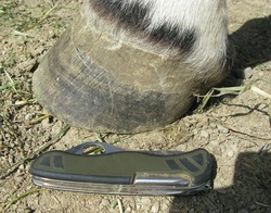
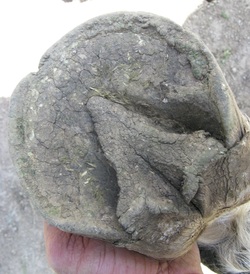
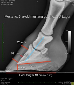
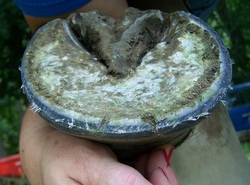
The three sets of images below compare the lateral and underside views, and the radiographs of the cadaver versus Mesteño's hoof. Note the different wear patterns in Mesteño's hoof due to the trim and the less abrasive limestone terrain.
As expected, the hoof radiograph for Mesteño shows a longer toe, greater digital breakover, higher heel and PA. These hoof characteristics are similar to those of my other young gelding (Nevada) and typical of domestic breeds. It is interesting that the HL zone in Mesteño's hoof is wider (20/18 mm) than the corresponding width generally observed in light breeds (15/15 mm) (refer to discussion above). The slight narrowing of the HL zone at the distal end (18 mm) is probably due to rasping of the hoof wall, or the absence of a radio-opaque marker to clearly delineate the hoof wall.
A more realistic comparison between the hooves of wild and domesticated mustangs would require tracking the history of a mature mustang recently captured and relocated to this area. Conversely, a domesticated horse could be released and followed in the wild to learn more about the relative contributions of environment and genetics to hoof conformation.
|
LF lateral and solar views of Mesteño's hoof. Pocketknife is about 4 in. long. Hoof length from toe to point where heel contacts ground surface is ~5 in. The width of the hoof at its widest part is ~5.5 in. Photographs taken about 4 weeks after conventional pasture trim. Compare to corresponding views of LF cadaver foot below. Photos by GA Lager
|
Wear pattern of sole and frog reflects grazing in rocky, limestone pastures.
|
_Comparison of digital X-ray radiographs of cadaver foot and 3-year
old domesticated mustang foot (Mesteño). Radiograph of domesticated foot, Dr. James Beckman, Gas Light
Equine. Image processing, annotations and measurements by GA Lager,
OsiriX for Mac OS X.
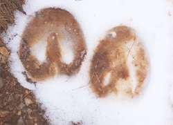
The load on the mustang hoof is shared by hoof wall, sole, bars and frog, reducing the load on the hoof wall (see image of barefoot hoof prints below). The advantage of this design is that it promotes blood flow in the hoof and provides more support for the foot with less stress on the laminae.
The loading of the hoof wall, due either to the nature of the trim, or the use of shoes, is sometimes referred to as "peripheral loading", an idea based on the research of Dr. Robert Bowker at Michigan State University. For more information on the advantages of natural hoof care, see the special report "Is Barefoot Better?", or check out Pete Ramey's new book, "Care and Rehabilitation of the Equine Foot". You might also be interested in the article "Why use the Wild Horse Model?"
Refer to our new web page on Natural Hoof Care for a PowerPoint slideshow and videos of the natural barefoot trim using both conventional farrier tools and the power (electric) grinder. Some images of Mesteño's hooves taken just after a natural trim are shown below. Compare the different views to those shown above for the pasture trim.
Mesteño's hooves after natural barefoot trim showing frontal view, and oblique view of RF solar surface. RF has a slight medial-lateral imbalance with more wear on the inside of the hoof. The greater length on the outside of the hoof causes flaring and cracking of the hoof wall. Note the small toe crack located just to the right of center in both views.
Lateral view RF. Toe shortened and heel height reduced relative to conventional pasture trim above.
Oblique and perpendicular views of LF solar surface. Good view of the mustang roll (bevel) in image on left. Load on hoof shared between hoof wall, sole, bars and frog.
For an alternative view, go to www.nanric.com/thewildhorsesfoot.asp, where Dr. Ric Redden discusses some of the reasons why the wild horse model should not be applied to domestic breeds.
TheHorse.com has also recently published an interview with Dr. Debra Taylor, Auburn University, who presented another veterinarian's view of barefoot versus metal shoes at the 2013 Western Veterinary Conference. Also refer to her video presentation "Is the hoof smart?: adaptability of the equine foot" for more information on how the foot responds to environmental factors. More on the anatomy and function of the hoof from a holistic perspective can be found on Dr. Tomas Teskey's facebook site.
TheHorse.com has also recently published an interview with Dr. Debra Taylor, Auburn University, who presented another veterinarian's view of barefoot versus metal shoes at the 2013 Western Veterinary Conference. Also refer to her video presentation "Is the hoof smart?: adaptability of the equine foot" for more information on how the foot responds to environmental factors. More on the anatomy and function of the hoof from a holistic perspective can be found on Dr. Tomas Teskey's facebook site.
Barefoot horse prints, front (left ) and hind. Note the contact area on the snow.
http://en.wikipedia.org/wiki/Horse_hoof
http://en.wikipedia.org/wiki/Horse_hoof
Copyright George Lager, 2011-2024. All Rights Reserved (®)
