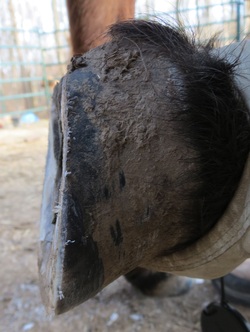
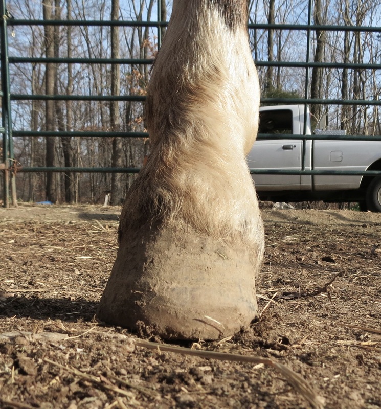
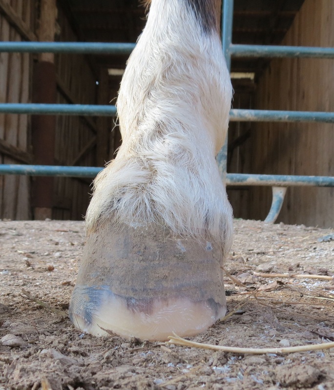
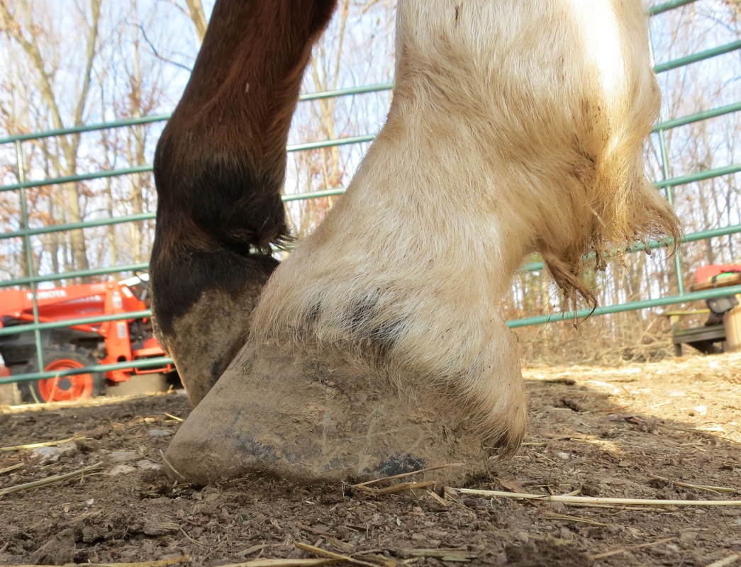
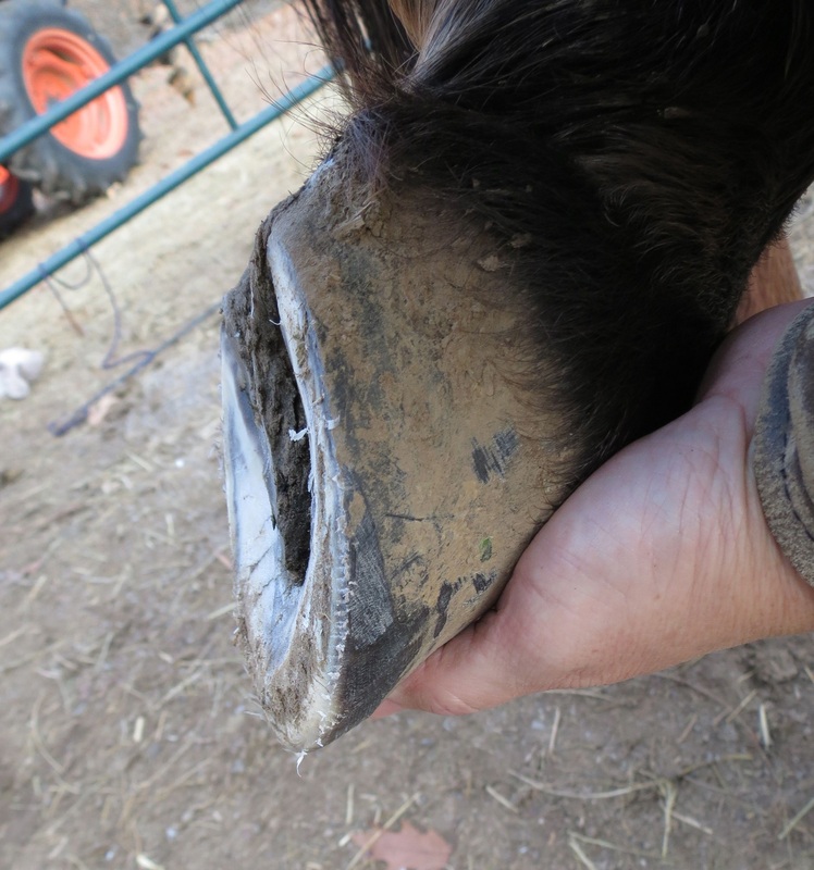
Natural Trim Based on the Wild Horse Model (Stephanie Ohlemacher and George Lager)
Natural Trim and Nutrition Clinic, Spring Lake Farm, Elizabeth, Indiana 09.26.2014. Photos by Diane Probst
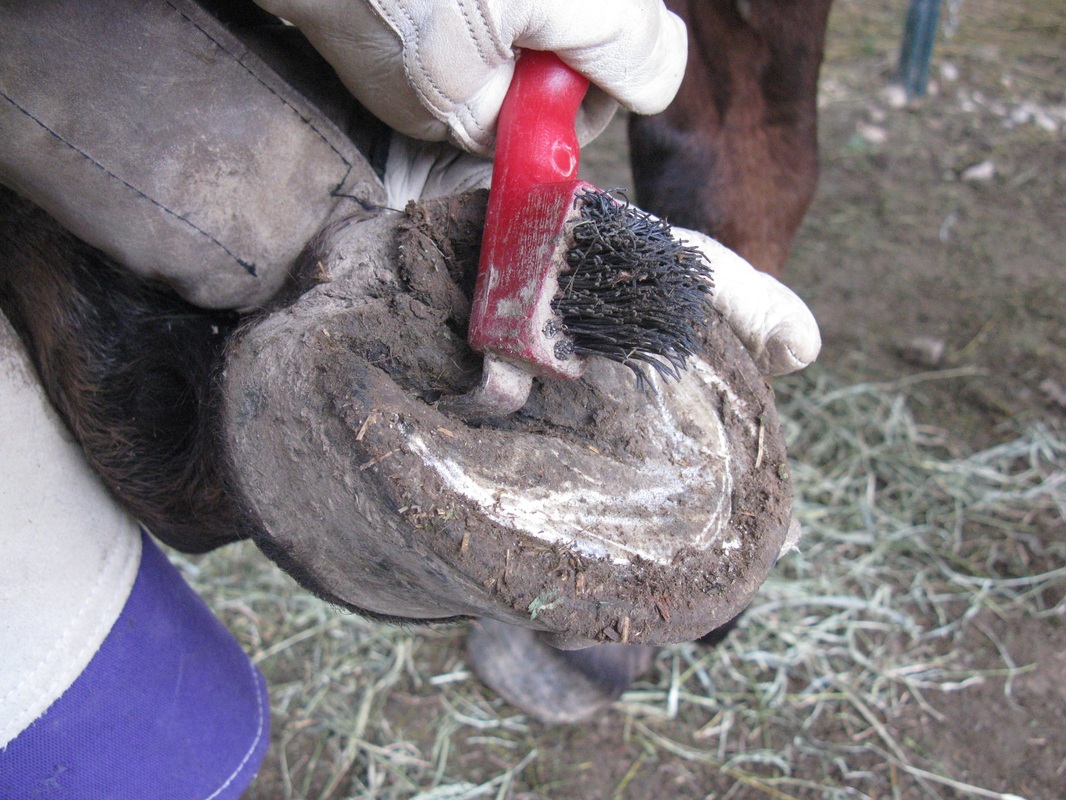
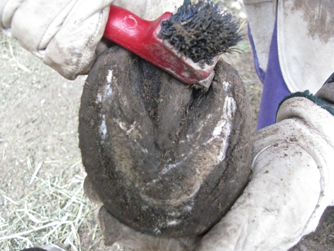
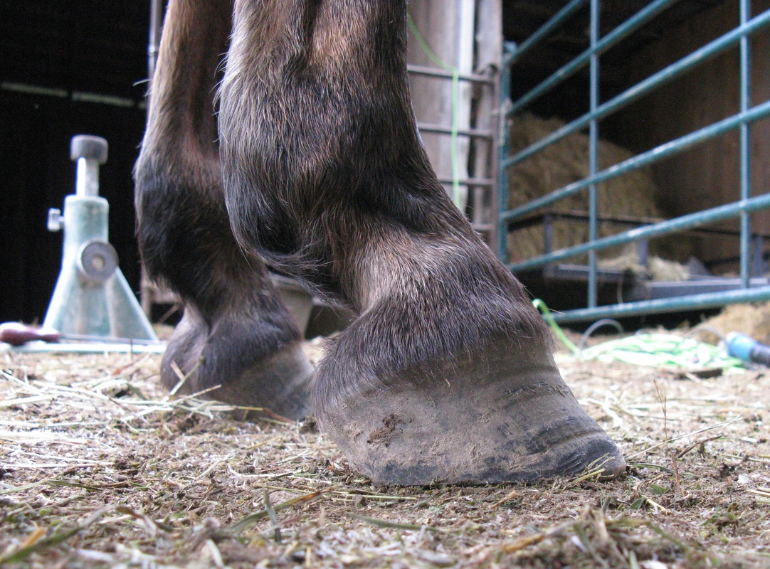
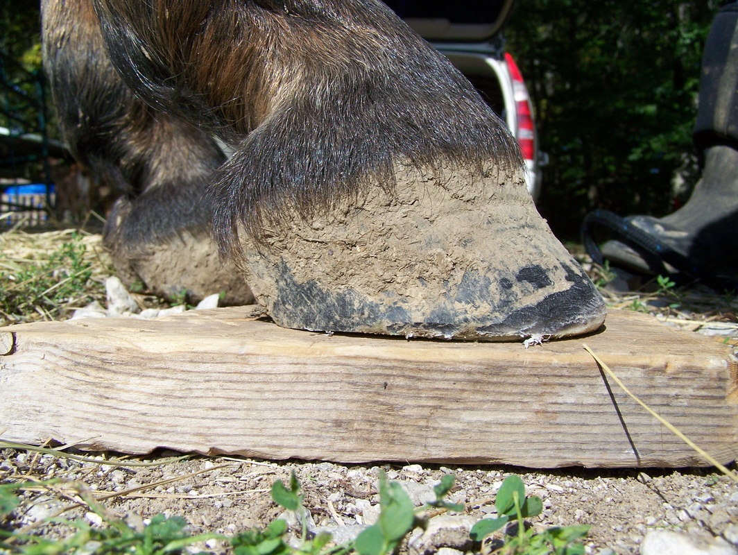
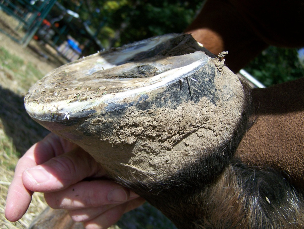
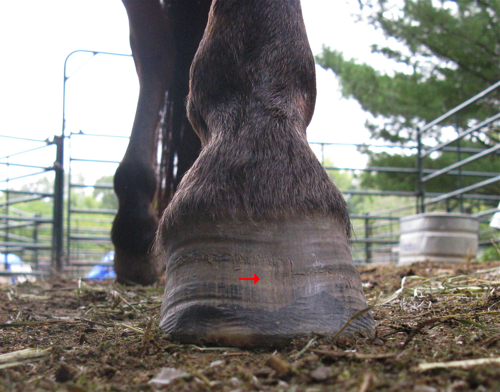
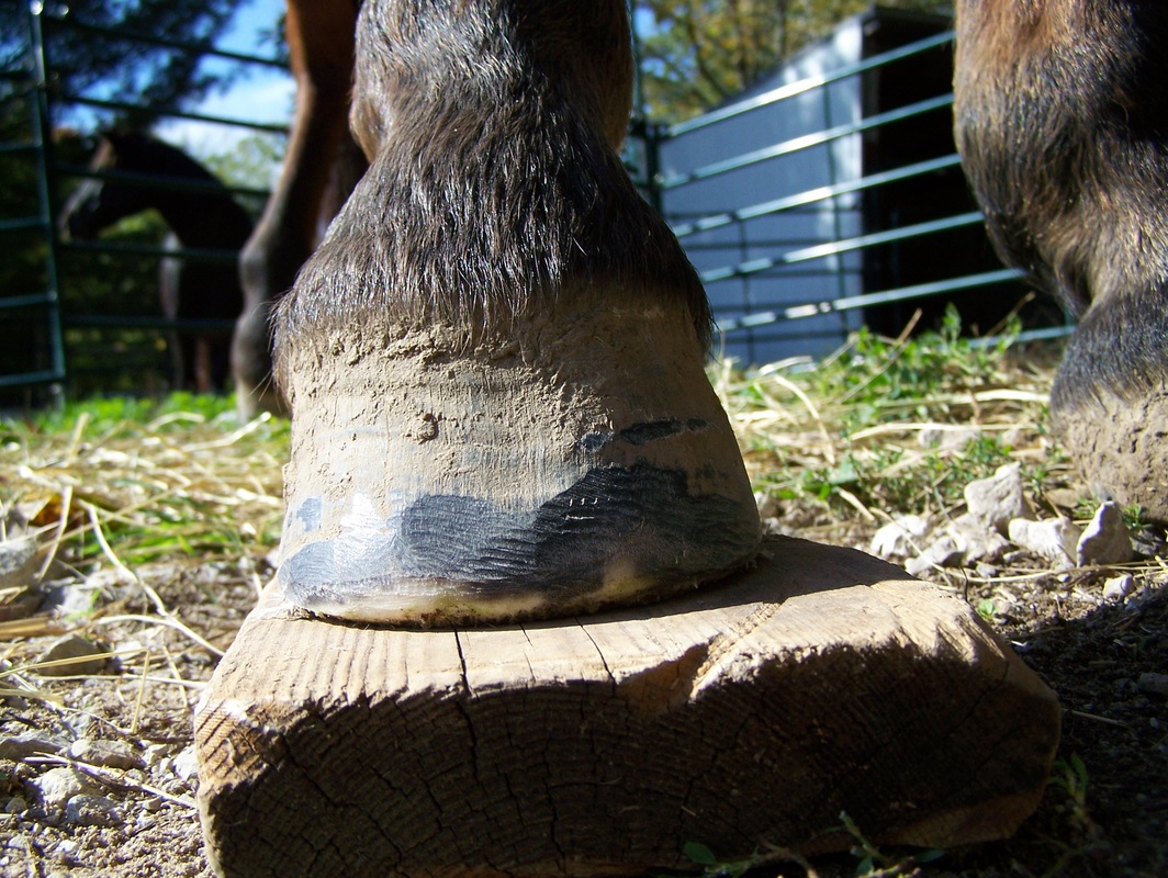
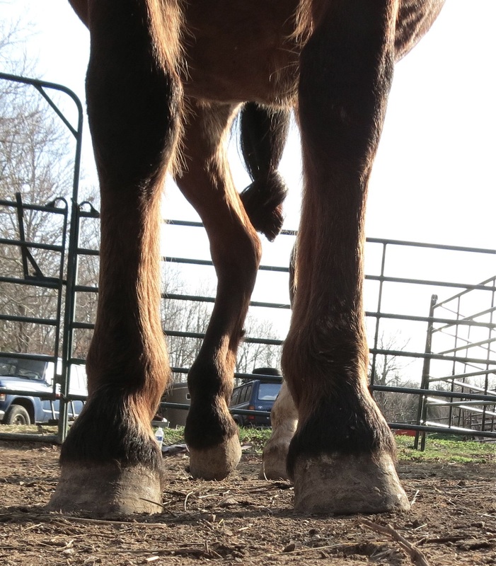
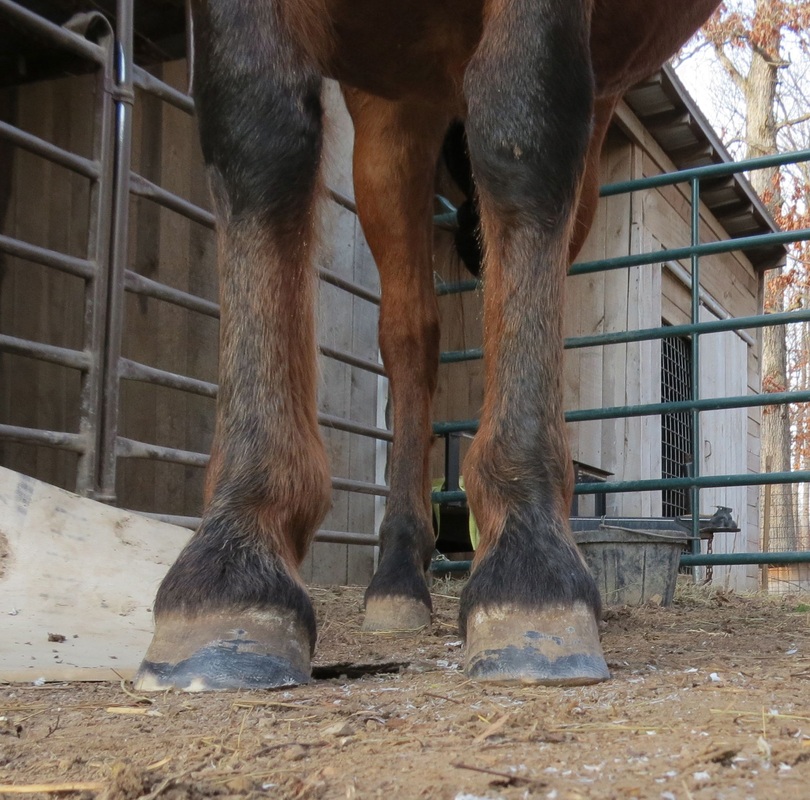
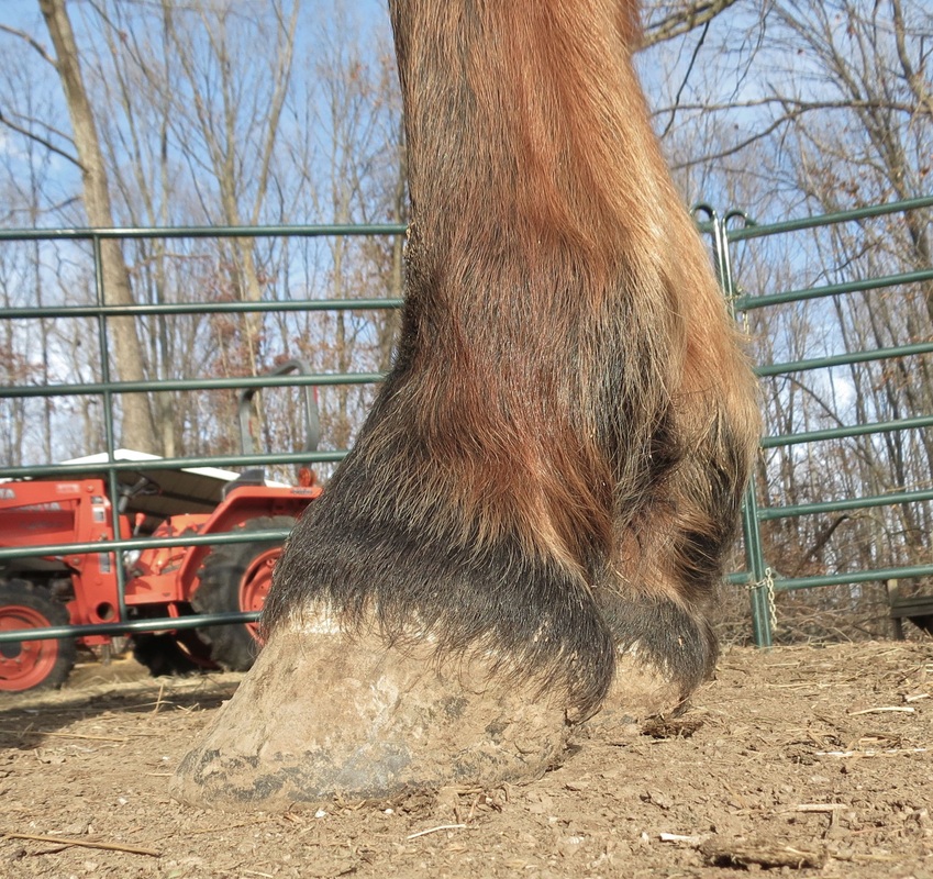
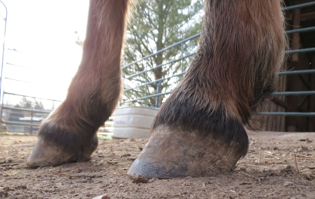
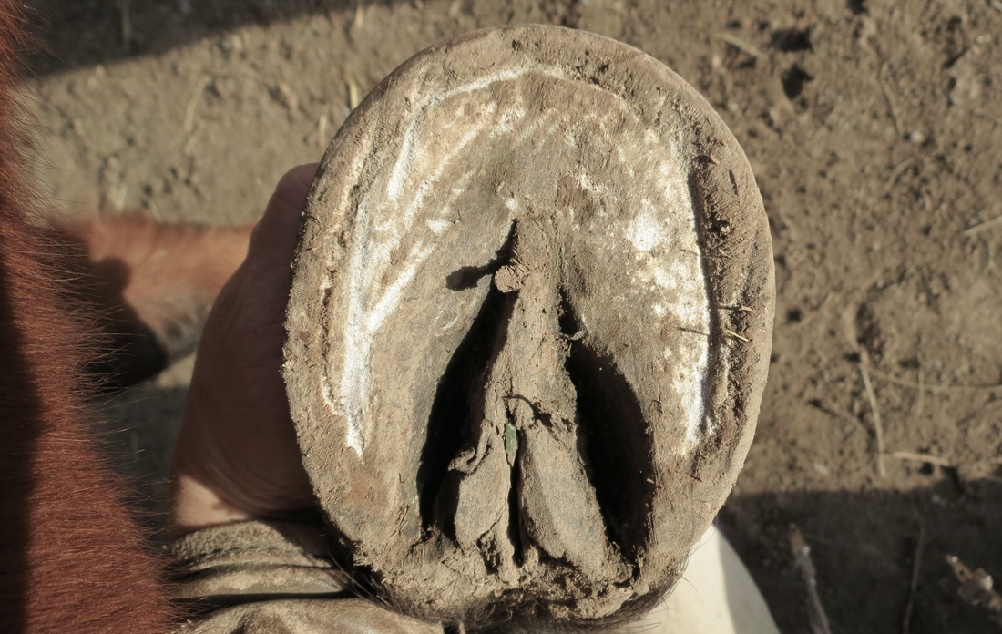
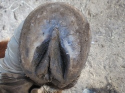
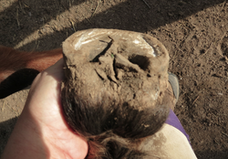
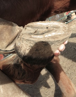
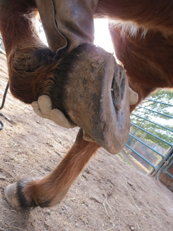
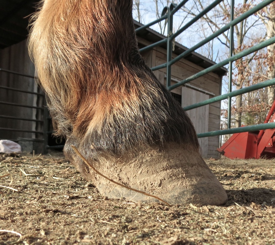
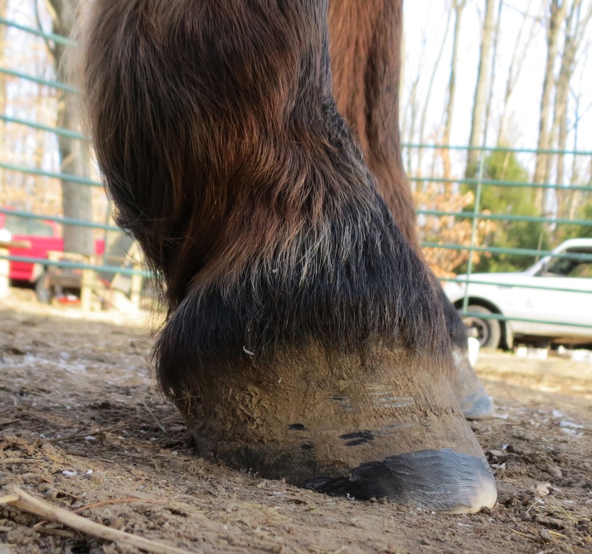
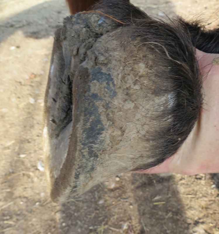
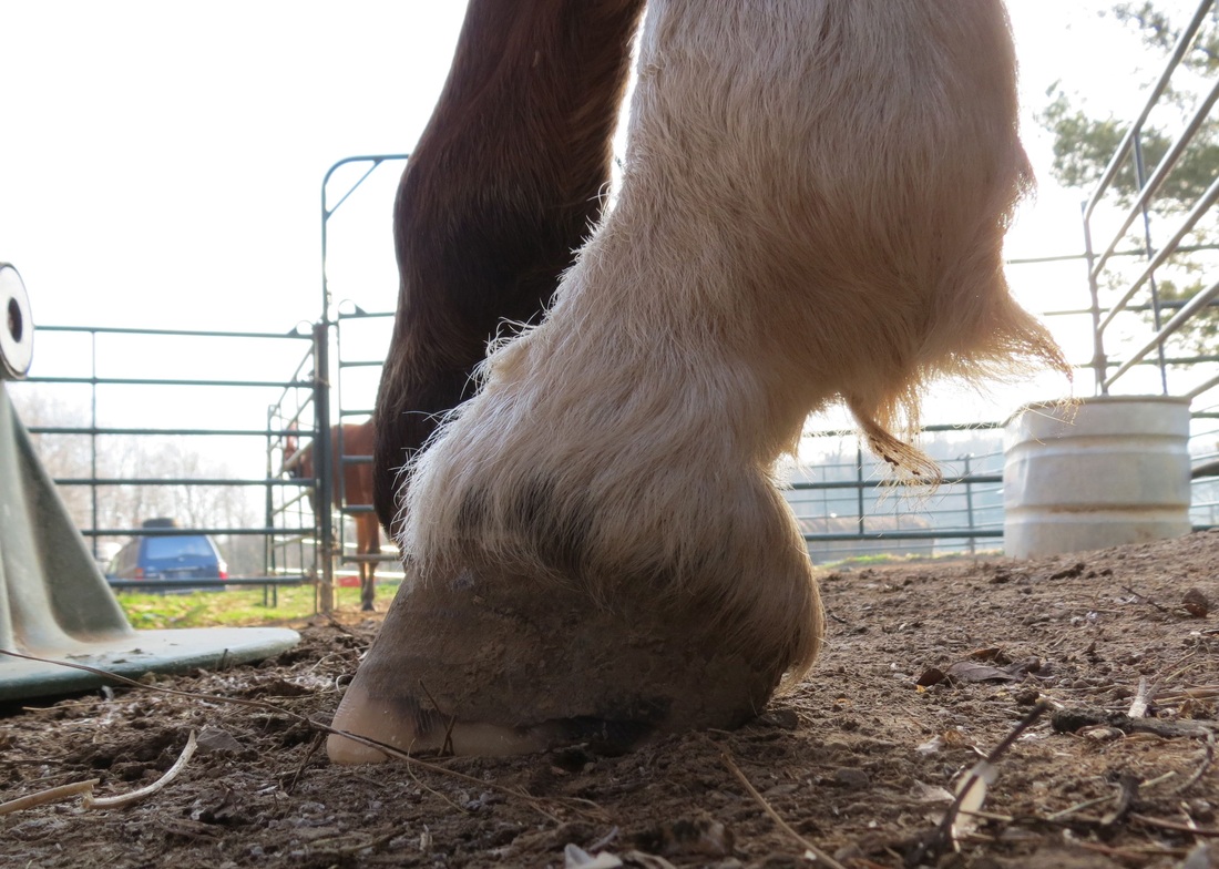
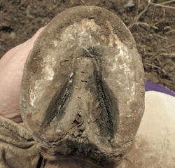
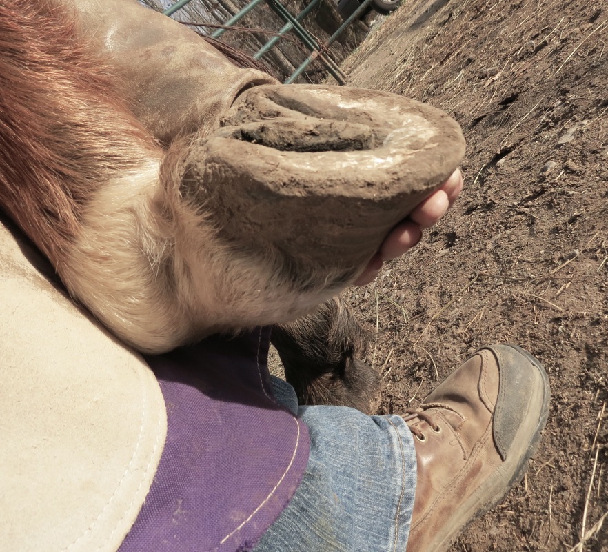
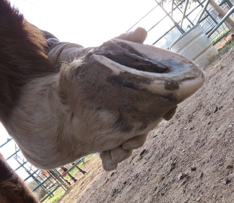
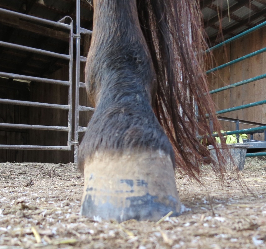
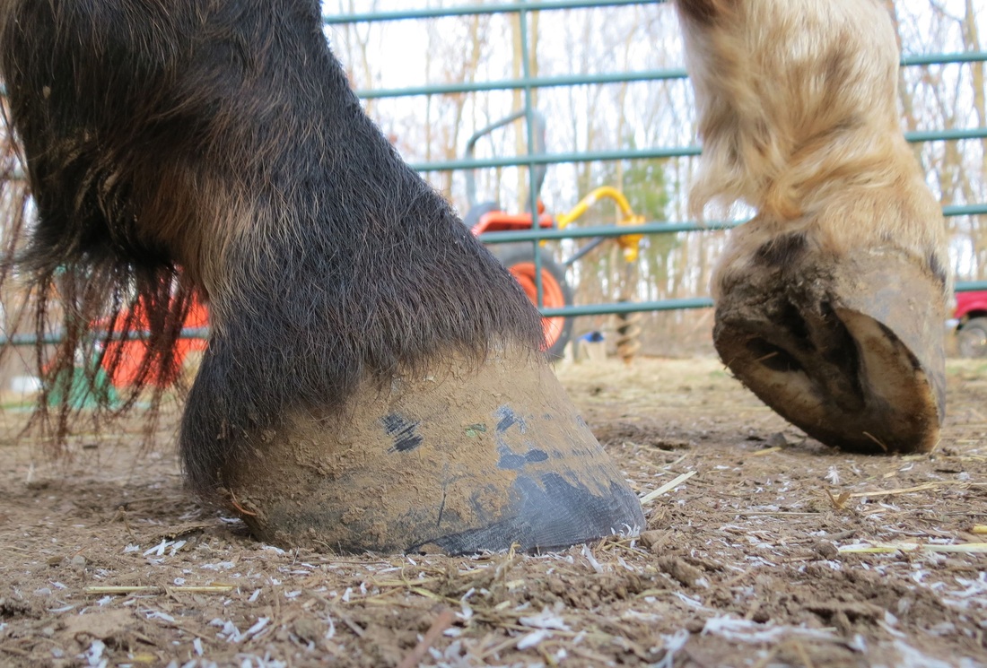
One important element that has been missing from our program of natural horse keeping is the natural (barefoot) trim based on the wild horse model. Most farriers in this area are advocates of the pasture trim, which loads the hoof wall relative to the sole, bars and frog. The toes are typically longer with greater breakover and the heels are higher, increasing the palmar angle. Refer to my article "The Mustang Hoof" where the wild horse hoof is compared to the hoof of one of our domesticated mustangs with a pasture trim.
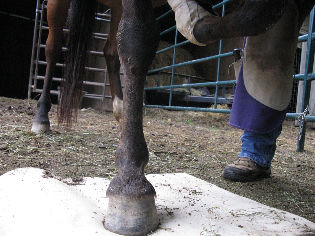
Our mustangs have never been shod (with the possible exception of the two older rescues with unknown history) and made the transition to the natural trim without problems. They regularly run in rocky limestone pastures and on a graveled paddock that partially encircles the pasture (Fig. 1). Because movement is so important to the proper functioning of the equine foot, the graveled paddock will be extended to encircle most of the main pasture ( à la Jamie Jackson’s Paddock Paradise). In most cases, our mustangs have 24/7 access to both pastures and paddocks.
Our mustangs have never been shod (with the possible exception of the two older rescues with unknown history) and made the transition to the natural trim without problems. They regularly run in rocky limestone pastures and on a graveled paddock that partially encircles the pasture (Fig. 1). Because movement is so important to the proper functioning of the equine foot, the graveled paddock will be extended to encircle most of the main pasture ( à la Jamie Jackson’s Paddock Paradise). In most cases, our mustangs have 24/7 access to both pastures and paddocks.
Fig. 1. The gravel road back to our main barn, which also serves as a paddock, will be extended to encircle the main pasture on the left.
On this web page, Stephanie Ohlemacher (S.O. NATURAL HOOF CARE, Elizabeth IN, retired) provides an introduction to the philosophy of the natural trim and some practical examples for those interested in learning more about this trimming method. Also check out the references in my article "The Mustang Hoof", in particular Pete Ramey’s web site and his new book on The Care and Rehabilitation of the Equine Foot, and the numerous other resources available in book form and on the Web (e.g., see above resources from S.O. NATURAL HOOF CARE). For a good example of how the barefoot trim affects hoof morphology, see the recent article by Clayton et al. (2011) in the Australian Veterinary Journal. The study includes quantitative measurements illustrated with both hoof photographs and X-ray radiographs over a 16-month period.
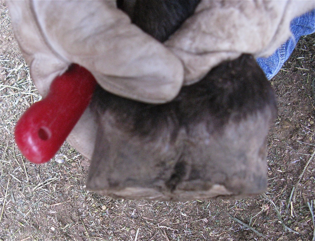
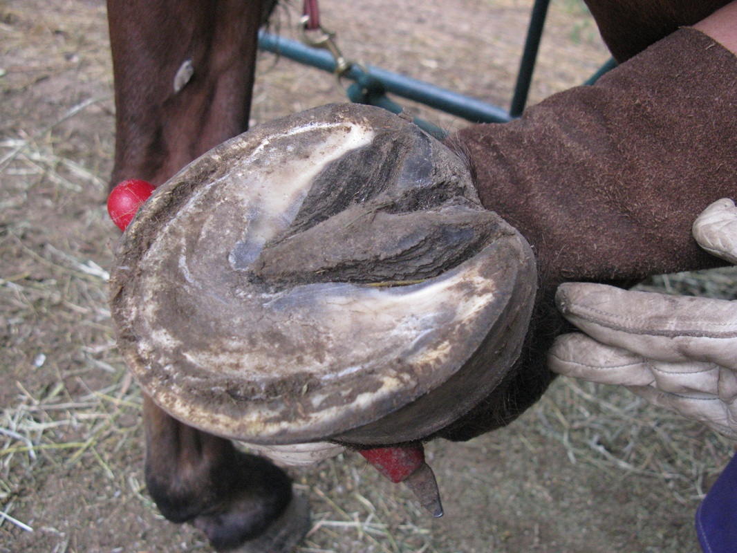
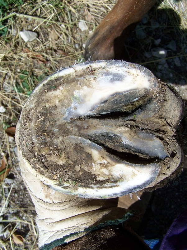
The slideshow below illustrates the natural trim with conventional tools for our 12-year-old rescued mustang gelding Comet, whose hind feet had not been trimmed in three years. Click on title page to view slides.
The slideshow below illustrates the natural trim with conventional tools for our 12-year-old rescued mustang gelding Comet, whose hind feet had not been trimmed in three years. Click on title page to view slides.
In the two videos that follow, Stephanie Ohlemacher uses the power (electric) grinder to trim Nevada, our 9-year old mustang gelding adopted through the Bureau of Land Management (BLM) program. Nevada was trimmed about 4 weeks ago. This is his second trim with the power grinder.
Effect of barefoot trim on hoof and limb deformities: case study of our mustang mare Montello (Stephanie Ohlemacher and George Lager)
The primary reason for this study is to determine if we can correct at least some of Montello's hoof and angular limb deformities and improve the soundness of her feet using barefoot trimming methods. It will be instructive to learn if this goal can be achieved over time on an older horse (15 years of age) diagnosed with chronic laminitis. This page will be updated with photos of her hooves after selected trims and a new set of X-ray radiographs on an annual basis.
Montello’s History
Very little is known of Montello’s history other than that she was adopted as a “yearling” in 2001 at a BLM adoption event in Edinburgh, Indiana and abandoned in early gestation in 2008 near Central Barren, Indiana. She was eventually brought to the New Beginnings Refuge near New Salisbury, Indiana (recently closed). According to the proprietors, her front hooves were so deformed and neglected that a rough trimming was necessary before she could walk without difficulty. The refuge was seriously considering euthanasia based on an initial examination of her hooves.
She was a very bad cribber during her first year or so on our farm and was diagnosed with a case of heaves. She prefers to enter the stall when fed and some situations, e.g., severe weather, can be stressful for her outside the stall. Based on her medical profile and behavior, our guess is that Montello was stalled and used as a broodmare for most of her life.
Montello's front hoof deformities are the result of chronic laminitis, based on the nature of the deformity and X-ray radiographs taken two years after adoption, i.e., greater width of horn-laminar zone at distal end and large palmar angle (~25°) (see 2010 photos and radiographs below). In addition, she has a conformational defect in her front knees (video below).
The end result is that she walks on the outside of her front hooves with very little wear on the inside. It would be very unusual for the BLM to offer a wild mustang for adoption with serious hoof or knee deformities. Most horses with major deformities are culled from the herds prior to adoption. It is possible that Montello had an underlying genetic predisposition, which was aggravated by poor hoof care and diet after adoption. For example, excessively high heels on both front and hind feet suggest very deep bedding and/or manure in stall (toe "sinks" into stall floor covering allowing excessive heel growth).
Since foaling, she usually has 24/7 access to pasture (fescue grass). Hay consists primarily of mixed grasses with some alfalfa, which is not supplemented by grain, or commercial horse feed. All our mustang horses are given a powdered vitamin/mineral supplement mixed with non-molasses beet pulp once daily. Free-choice salt and salt blocks are always available.
As far as possible, we make every attempt to mimic the grazing environment of mustangs in the wild, e.g., non-cultivated pastures with no weed control other than mowing. Pastures are rotated and fertilized with composted horse manure.
Montello has not shown any signs of obvious lameness in any of her feet since arrival at our farm in 2008. As noted above, she has recently made the transition from the pasture trim to the natural barefoot trim with no adverse effects.
X-ray radiographs and photos of front feet (09.18.2012)
All radiographs of pasture trims were taken about 6 weeks after trim. For barefoot trims, radiographs of pre- and post-trims were taken during the same farm call (Dr. James Beckman, Gas Light Equine). The barefoot pre-trim represents about 4 weeks of hoof growth since the first barefoot trim. Position of frog indicated by red arrow in radiographs of pasture trim. For barefoot trim, thumbtack placed slightly off apex of frog.
Dorsopalmar (DP) radiographs of pasture trim are digitized images of film radiographs (films obtained by Dr. Sharon Christie, Smith Animal Clinic and digitized by Mr. Dwight Fredericks, Harrison County Hospital). Lateral digital radiographs of pasture trim recorded by Gas Light Equine. Image processing and annotations by GA Lager using OsiriX software package for Macintosh OS X.
Because of time constraints related to sedation, trims were completed quickly using both external hoof guides and pre-trim radiographs. Note that the optimal conformation cannot be achieved in one trim for fear of lameness. Further adjustments to toe length and heel height based on post-trim radiographs and external guides were made in followup trim on 10.06.2012 (see below).
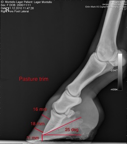
Left front (LF) lateral radiographs and photos from left to right: pasture trim 08.26.2010 (radiograph 11.12.2010); barefoot pre-trim 09.18.2012; barefoot post-trim 09.18.2012.
|
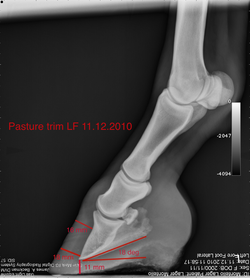
Six weeks after pasture trim LF; two years (2010) prior to barefoot trim; high heels resulting in larger angle between P3 solar margin and ground surface (palmar angle-PA); width of horn-laminar (H-L) zone slightly greater at distal end; mild coffin bone (P3) remodeling at tip ("ski tip"); misalignment of natural harmonic curve
|
Four weeks LF after first barefoot trim; two years prior pasture trim (2010-2012) with excessively high heels have caused greater degree of remodeling at tip pf P3 and pronounced flaring of dorsal hoof wall near toe; lower heel height improved alignment of natural harmonic curve, lowered PA relative to pasture trim, better P3-P2 alignment
|
Second barefoot trim with lower heel height has further reduced PA; relaxed hoof wall reflected in greater width of H-L zone at proximal end; reduced flaring; sufficient sole depth beneath tip of P3 (~11 mm); digital breakover ~23 mm; toe left longer to assert slight lever force, allowing tendons and ligaments to adapt to lower heel height and inhibiting potentially severe side effect of "knuckling over" due to tight
tendons and muscles from long-term high heels |
Comparison of LF lateral views before and after barefoot trim. For post-trim, shorter toe and lower heel; flare reduced on hoof wall near toe.
Right front (RF) lateral radiographs from left to right: pasture trim 08.26.2010 (radiograph 11.12.2010); barefoot pre-trim 09.18.2012; barefoot post-trim 09.18.2012
|
Similar issues to LF but to a greater degree; greater PA and more significant misalignment of harmonic curve
|
Better P3-P2 alignment four weeks after first barefoot trim; lower PA; sole depth beneath P3 unchanged relative to pasture trim; pronounced flaring of dorsal hoof wall; increased remodeling of P3 after two years of pasture trim
|
Further reduction in PA with lower heel height; P3-P2 angle reduced by several degrees with decease in PA; lower heel height resulted in relaxation of soft tissue at back of foot, enhancing heel expansion and proper hoof mechanics; as for LF, toe left longer to assert slight lever force
|
Comparison of RF lateral view before and after barefoot trim. Adjustments similar to LF. More flaring on RF vs LF hoof wall near toe. Note that pre-trim photo (l) not true lateral.
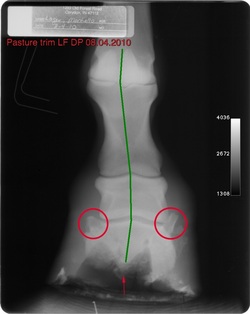
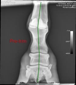
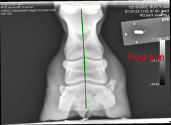
LF dorsopalmar (DP) (anterior-posterior) radiographs and photos from left to right: pasture trim 06.18.2010 (radiograph 08.04.2010); barefoot pre-trim 09.18.2012; barefoot post-trim 09.18.2012
|
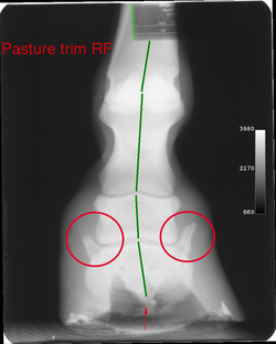
Excessively high heels six weeks after pasture trim; medial-lateral imbalance in leg; P3-P2-P1 misalignment (green line); excessive side bone lateral aspect (circled) formed in response to body's attempt to preserve natural alignment due to angular limb deformity
|
Slight improvement in P3-P2-P1 misalignment four weeks after first barefoot trim; frog better developed and closer to ground surface but deviated due to excessive load on lateral side; heels still high; soft tissue lateral to P2 more relaxed
|
After second barefoot trim improved alignment with P2 positioned correctly relative to side bones. Frog in contact with ground surface but still deviated.
|
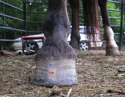
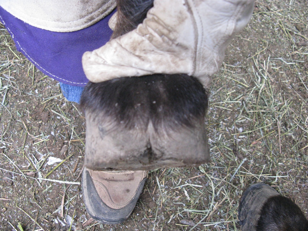
Dorsal view LF before and after barefoot trim. Flaring reduced on medial side (red arrow). Note recessed area and curved growth lines on hoof wall due to a number of physiological factors associated with unhealthy foot.
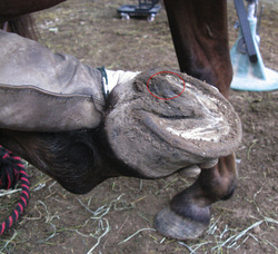
Oblique solar (bottom) view LF before and after barefoot trim. Trimmed excess bar height compressing sole and elevating hairline medially (red circle). Compare pre-trim solar view to pre-trim medial lateral view below.
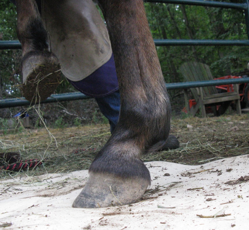
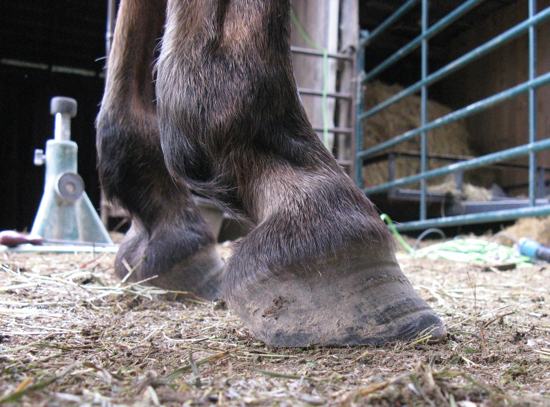
View from heel illustrating wall and heel height before and after barefoot trim.
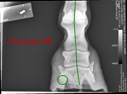
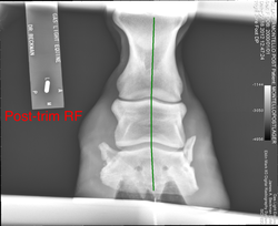
RF DP radiographs and photos from left to right: pasture trim 06.18.2010 (radiograph 08.04.2010); barefoot pre-trim 09.18.2012; barefoot post-trim 09.18.2012
|
Six weeks after pasture trim heel height and foot and leg deformity RF similar to LF; greater deviation of frog (red arrow); more pronounced P3-P2-P1 misalignment
|
Better P3-P2-P1 alignment four weeks after first barefoot trim; note height of frog above ground surface relative to pasture trim; medial-lateral imbalance; bone loss P3 solar margin lateral side (circle) due to excessive load on outside of foot
|
After second trim, further adjustments in heel height and toe length over time will be required to improve alignment and medial-lateral imbalance
|
Dorsal view RF before and after barefoot trim. Recessed area on hoof wall enlarged relative to LF with greater curvature of growth lines (red arrow).
Oblique solar view before and after barefoot trim.
View from heel illustrating hoof wall and heel height before and after barefoot trim.
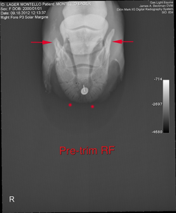
LF and RF radiographs P3 solar margins barefoot pre-trim 09.18.2012; flatter distal margin of P3 indicates slight bone loss (flatter margin parallel to line drawn between red points); bone loss more severe for RF; P3 side bones (lateral cartilage calcification) for RF consistent with more severe deformity (red arrows).
Followup hoof trim 3 weeks post X-ray study
As noted above, barefoot trims on 09.18.2102 were based on both X-ray radiographs and external hoof guides because of time constraints related to sedation. In this series of hoof trims (10.06.2012), the above post-trim radiographs were used to make further adjustments (primarily, toe length and heel height) to the front hooves. As illustrated below, these adjustments resulted in significant improvements in both hoof and leg conformations.
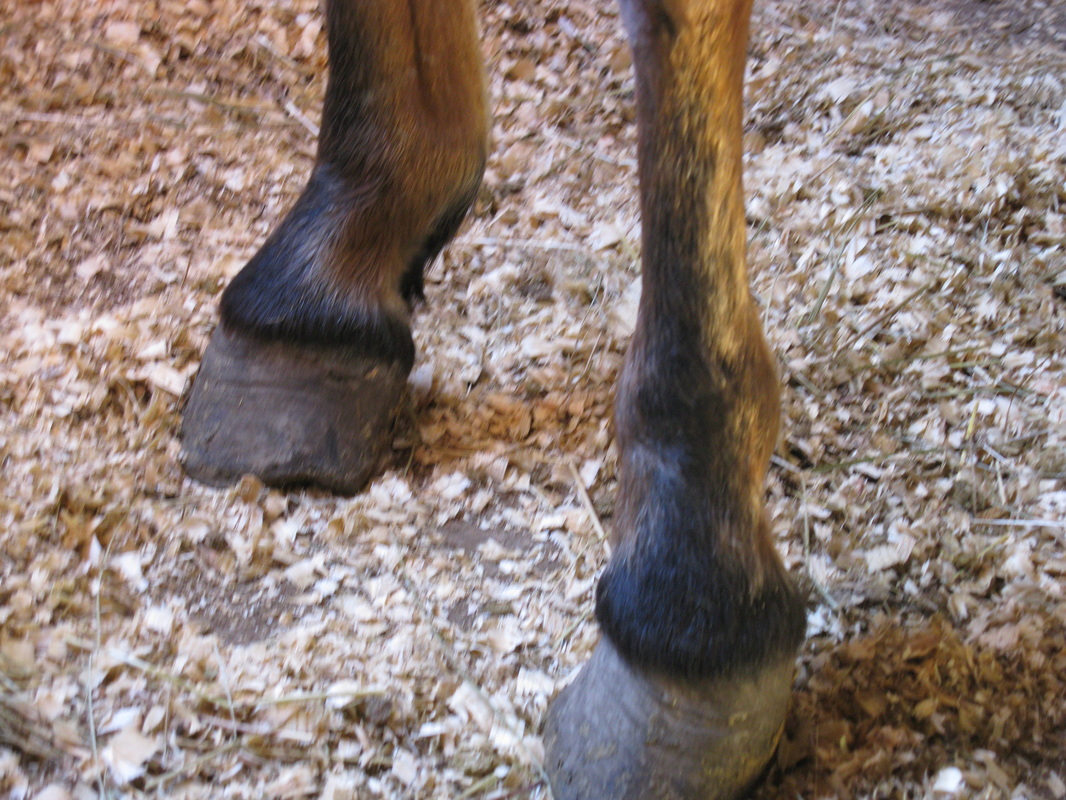
Far left: oblique view of front hooves after rough trim to enable mare to walk without difficulty; photographed at rescue facility (11.10.2008)
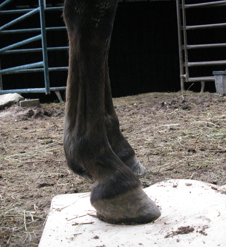
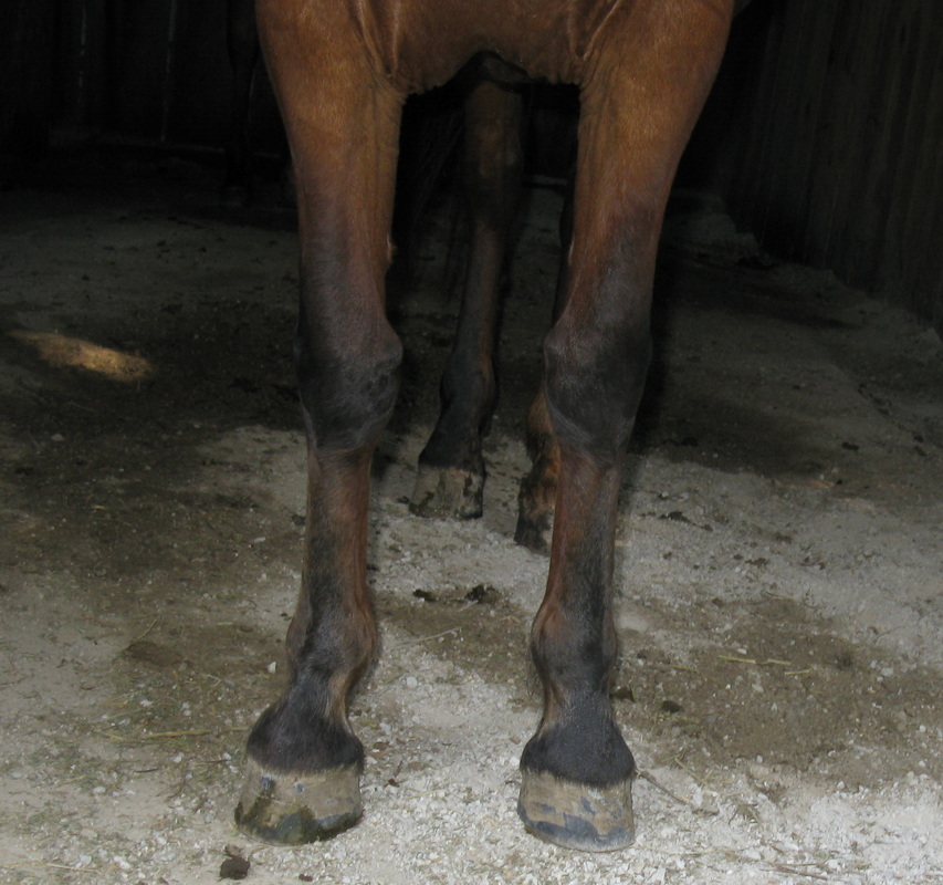
Center: anterior view of hooves and knees showing angular limb deformity (knock-kneed, or carpus valgus); toe shorten to correct medial-lateral hoof imbalance but heels left high (pasture trim, 12.14.2008)
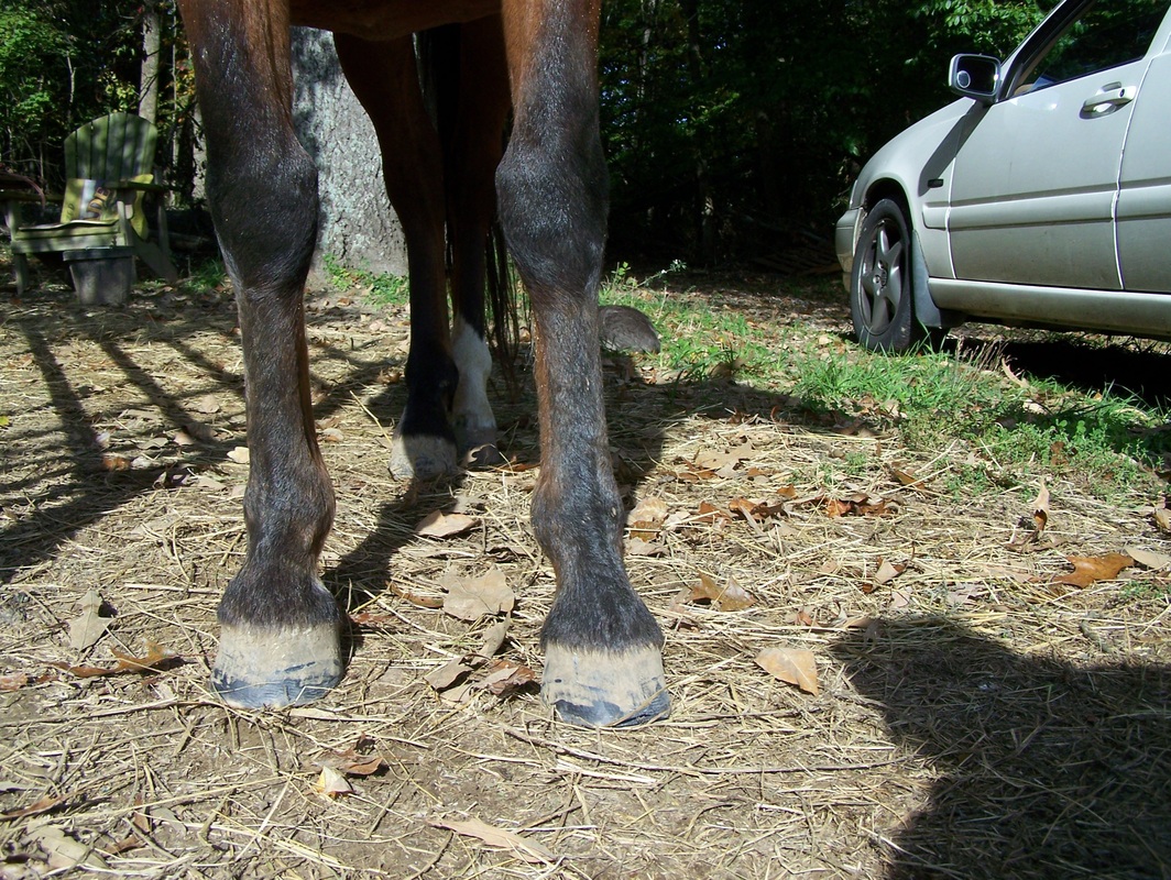
Right: anterior view after barefoot trim 10.06.2012 using X-ray radiographs as guide; shorter toe and lower heel with less angular deviation of knees
Far left: lateral view LF at rescue facility
Center: lateral view LF after barefoot trim 09.18.2012; flare on dorsal hoof wall
Right: lateral view after barefoot trim 10.06.2102; shortening and beveling of toe; flare reduced; lower heel height, resulting in further relaxation of hoof wall (more uniform width H-L zone and lower PA)
Center: lateral view LF after barefoot trim 09.18.2012; flare on dorsal hoof wall
Right: lateral view after barefoot trim 10.06.2102; shortening and beveling of toe; flare reduced; lower heel height, resulting in further relaxation of hoof wall (more uniform width H-L zone and lower PA)
Solar view LF trim 09.18.2012 (l) vs trim 10.06.2012. Hoof wall 09.18.2012 trim slightly higher at toe to alleviate pressure on sole.
Lateral view RF trim 09.18.2012 (l) vs trim 10.06.2012; flare reduced, resulting in tighter hoof wall connection.
Solar views RF trim 09.18.2012 (far left) vs trim 10.06.2012 (center and right); bars and heels reduced in height; weight-bearing surface of heels moved to posterior area of foot as heel height lowered, increasing and elongating solar surface area; removed diseased and tattered frog tissue; toe further shortened and beveled; toe left longer until heel height stabilizes between successive trims
Dorsal view RF trim 09.18.2012 (l) vs trim 10.06.2012
Barefoot trim 12.03.2012
Pre-trim photos represent about 2 weeks (17 days) of growth since last trim. Hind feet, which are very upright with excessively high heels, are also included in this installment.
Further adjustments in heel height and toe length over the past three months have changed wear pattern on front hooves. In particular, solar views show wear on the medial side of hoof, i.e., she no longer walks almost entirely on the outside of hoof. Solar concavity more developed except for flattened area near toe. Refer to the article on The Mustang Hoof for photos and X-ray radiographs illustrating solar concavity in the wild horse hoof.
Further adjustments in heel height and toe length over the past three months have changed wear pattern on front hooves. In particular, solar views show wear on the medial side of hoof, i.e., she no longer walks almost entirely on the outside of hoof. Solar concavity more developed except for flattened area near toe. Refer to the article on The Mustang Hoof for photos and X-ray radiographs illustrating solar concavity in the wild horse hoof.
Anterior view of front hooves and legs before and after trim
Flares present on lateral side RF and medial side LF. Flattened sole near toe weight-bearing surface on LF. Refer to comments below.
Left front before and after trim
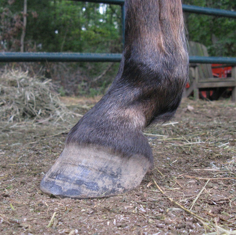
Lateral view LF before and after trim. Note lower heel height in trimmed hoof.
Solar views LF before and after trim. More wear seen on the medial side in latest pre-trim. Prior to barefoot trim wear was almost entirely on lateral side. More balanced load achieved by progressively lowering heel, shortening toe and reducing flares. Sole left essentially untouched, except for removal of chalky sole near apex of frog.
Heel views LF pre-trim 12.03.2012 (l) versus 09.18.2012. Note different wear patterns in pre-trim photos, i.e., less wear on medial side in 09.18.2012 trim. Linear shadow on medial side in 12.03.2012 trim marks location of lateral sulcus.
Oblique solar view LF before and after trim. Loose frog tissue in pre-trim shadowed in corresponding heel view above. Removed chalky sole near apex of frog revealing newly developed concavity. Flattened area (no concavity) near dorsal aspect of sole is at present weight-bearing surface (see also anterior view above). This area will become concave in shape with continued re-balancing of hooves and legs. Note backwards heel bevel in post-trim magnified image. Bevel mimics lower heel height to allow gradual lengthening of contracted tendons and muscles due to chronic high heels.
Right front before and after trim
RF lateral views before and after trim. Bevel less than LF because of thin wall at toe (see solar view below).
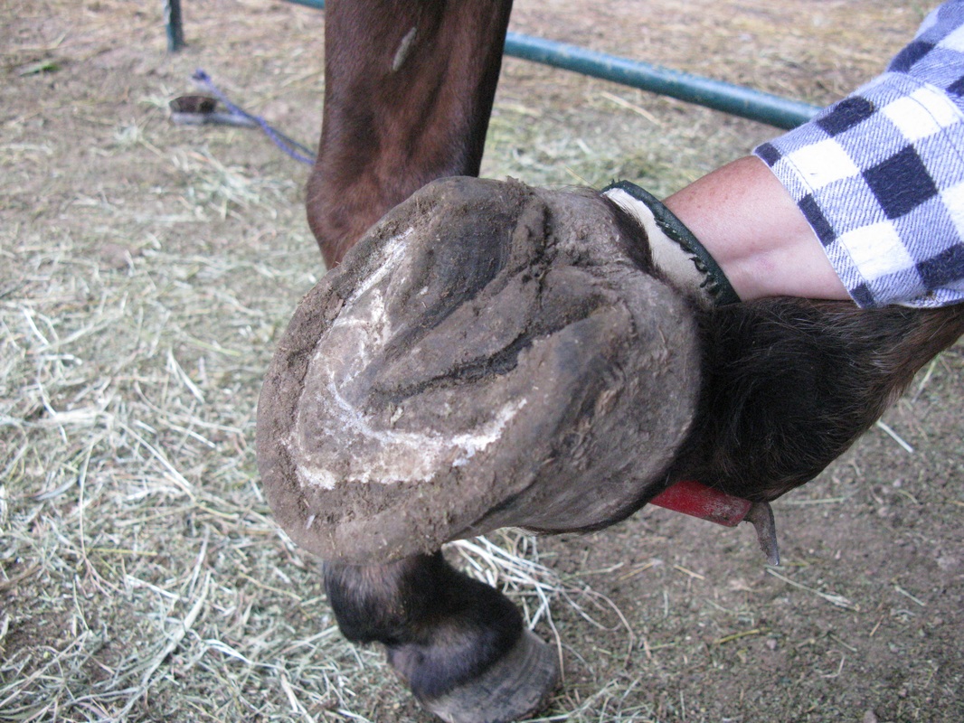
Solar views RF before and after trim. Poor hoof wall connection (separation along white line) until imbalance corrected and lateral flare grown out. Note thin wall at toe and increased wear on medial side of hoof. In this case, a normal amount of beveling at toe would intrude into live sole. Overturned bars on lateral side trimmed to sole level.
Side view RF before and after trim showing the development of concavity in sole. As in LF, flattened area near toe in post-trim.
Left hind before and after trim
Dorsal view left hind (LH) before and after trim. Steep hoof wall on hinds due to excessively high heels. See pre-trim video below for abnormal movement of LH which becomes less noticeable in post-trim video.
Lateral view LH before and after trim. Lower heel and shorter toe length after trim.
Solar view LH before and after trim.
Oblique solar view LH before and after trim. Beginning to develop good solar concavity post-trim.
Right hind before and after trim (with exception of dorsal and lateral views)
Dorsal view right hind (RH) after trim. Decrease in slope of hoof wall as heel lowered and toe shortened. Healthiest hoof on horse with less flaring and less medial-lateral imbalance.
Solar view RH before and after trim. Small radial cracks in sole post-trim developed in layer of chalky sole.
Oblique solar view RH before and after trim. As in LH, good sole concavity beginning to develop. Heel height close to optimal but flattened sole near toe still present.
Videos showing gait before and after trim
Gait not as smooth before trim. Note in particular the movement of the right front and left hind feet. Compare to video at beginning of section on case study.
Gait not as smooth before trim. Note in particular the movement of the right front and left hind feet. Compare to video at beginning of section on case study.
Smoother gait after trim. Less angular deviation of feet and knees.
Copyright George Lager, 2011-2019. All Rights Reserved (®)


