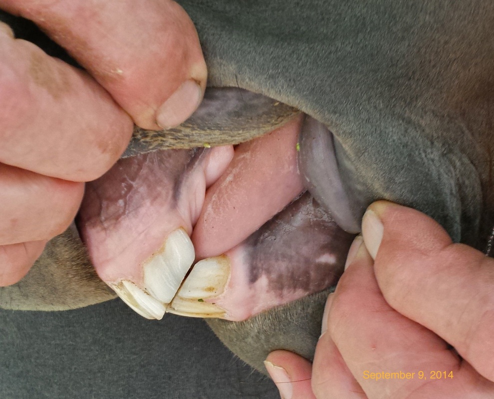
Dental Development of Our Mustang Filly, Morning Star
In a recent article on TheHorse.com, Dr. Jack Easley, a noted equine dentist in the Kentuckiana region, discussed the importance of monitoring the development and shedding of deciduous (baby) teeth in young horses. Deciduous teeth are shed at periodic intervals until the horse is about 5 years old, at which time the mouth has a full complement of permanent incisors and molars.
We decided to document the changes in Morning Star's mouth with images and observations recorded during each dental exam (equine dentists, Dr. Sharon Christie, Smith Animal Clinic; Dr. James Beckman, Gas Light Equine). This will be a good learning experience for me and, perhaps, for others as well. It will be interesting to see how natural horsekeeping affects the development of her mouth. We will update this page periodically after each exam.
For a basic introduction to equine dentistry refer to the numerous online resources, such as those provided by The American Association of Equine Practioners and by magazines for horse owners, such as The Horse and EQUUS. If you are interested in a more advanced treatment of topics in equine dentistry, a good choice is the book Equine Dentistry by J Easley, PM Dixon and J Schumacher, 2011, 3rd Ed. For a holistic approach to dentistry, in particular the importance of incisors, refer to the recent article by Spencer LaFlure in Equine Wellness.
Exam 1: April 21, 2011
Morning Star's first full mouth exam with a speculum was conducted this spring in April at 20 months of age. At this age, all incisors and premolars (excluding the first premolars, or wolf teeth) were deciduous. The first permanent molar (there are no deciduous molars) with an average eruption time of 9-12 months was also observed.
No unusual wear patterns were noted. The sharp enamel points that can form on the buccal (cheek) side of the upper premolars and lingual (tongue) side of the lower premolars were not sufficiently developed to warrant rasping. Images of the upper and lower incisors and premolars in two quadrants of the mouth are shown below.
|
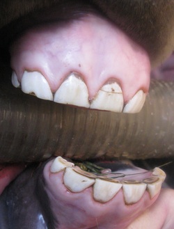
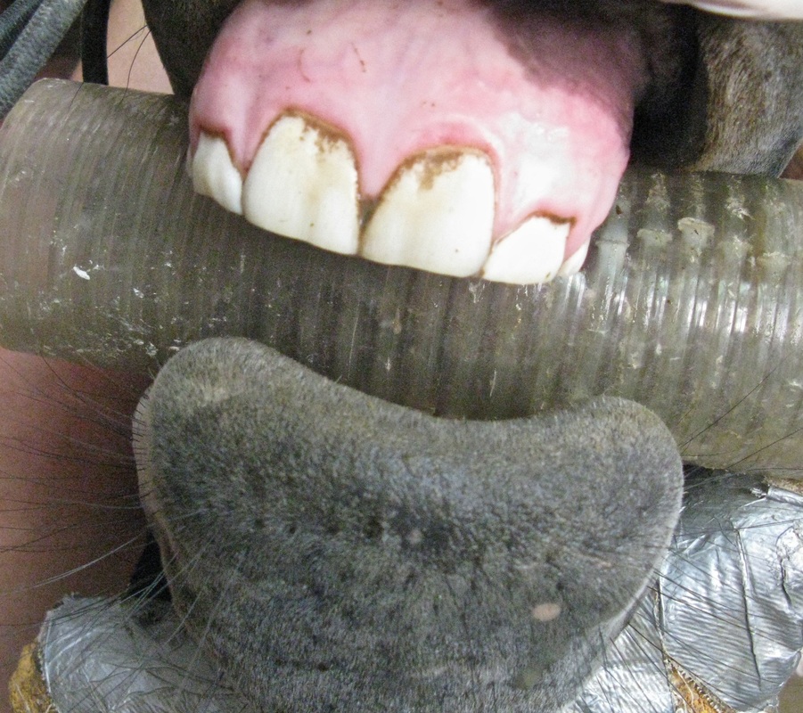
Image of the deciduous incisors. The oldest incisors (the two upper and lower central incisors) are shed at about 2.5 years. Note the different wear patterns of the central (Triadan 401), intermediate (402) and corner incisors (403) (refer to text). See figure below for Triadan numbering system. Photo by GA Lager
|
Image of the deciduous premolars and first permanent molar (209). Teeth 206-211 are collectively known as the cheek teeth. The smaller white tooth in front of 206 and displaced to the left (medially) is the first premolar, or wolf tooth (205), which does not have a deciduous precursor. The dental table, or occlusal surface, is not flat but inclined at ~ 20° to the horizontal plane. Occlusal angle decreases with age to a limiting value of 10-15° in adult horses. Photo by GA Lager
|
|
Teething bumps. About four months after the dental exam (08/28/2011), a teething bump (eruption cyst) was observed at the bottom of the mandible at the approximate position of the second premolar (406). The corresponding teething bump on the left side of the jaw developed a few weeks later. As of 11/26, both bumps have disappeared. Photo by GA Lager
|
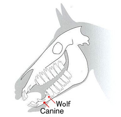
Arrangement of hypsodont, or high-crown teeth, in skull. Hypsodont teeth (i.e., incisors and cheek teeth) continue to erupt throughout life to compensate for wear. The canine and wolf teeth are brachydont (low crown) teeth, which are similar in anatomy to those in humans and other mammals.
|
Modified Triadan System for dental nomenclature
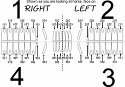
Modified Triadan System for dental nomenclature.
_ In some cases, only one number is used to represent each set of four teeth. For example, incisor number 101 can refer to a specific tooth, or to the set consisting of 101, 201,
301 and 401, starting with 101 in the upper right quadrant of the mouth and ending with 401 in the lower right quadrant. Not all sets of teeth occur in groups of four. For example, Morning Star has two small wolf teeth in the upper jaw (105, 205). Link to this table, which lists the average eruption times of deciduous and permanent teeth. Wolf teeth of horses and canine teeth in mares are vestigial structures, meaning that they no longer serve a function in the modern horse.
In earlier exams, the same dental nomenclature is used for both deciduous and permanent teeth. When there is some ambiguity, the tooth number will be prefixed by the term deciduous, or permanent.
In earlier exams, the same dental nomenclature is used for both deciduous and permanent teeth. When there is some ambiguity, the tooth number will be prefixed by the term deciduous, or permanent.
Incisors
_The magnified image of the lower incisors (below) clearly shows some
significant differences between the wear patterns of the central,
intermediate and corner incisors. For example, corner incisor (403) has a
deep tooth cup. That's because it's the youngest tooth and has been in
wear for the least amount of time. Also, note that only the labial
side (inner lip side) of this incisor is in wear.
_In
contrast, the cups in the two oldest incisors (401, 301) are
relatively shallow and surrounded by an elongated, oval-shaped structure called the
enamel ring. The brownish
linear structure on the labial side of the cups is the dental star.
In the drawing below, longitudinal (front to back, or sagittal) and transverse (across) sections of a lower central permanent incisor (301 or 401) are represented in terms of horse age (3, 5, 7 years, etc.). We will talk more about how to determine horse age from incisors in a future web post. This type of diagram is also useful in visualizing the effects of wear and anatomy on the appearance of the occlusal surface of deciduous incisors. Refer to this diagram and the caption in the discussion that follows.
Anatomy of equine permanent incisor. Longitudinal (or sagittal) and transverse cross sections of a permanent lower central incisor. Sections "cut" parallel to numbered dashed lines show wear patterns of occlusal surfaces in terms of approximate horse age. Labels: E, enamel (white); D, primary dentin (yellow); I, infundibulum (funnel-shaped, enamel fold, or infolding, at top of incisor, mostly filled with cement (C); the part at the top is unfilled, or filled with food particles ("cup"), lower portion filled with cement (enamel ring and enclosed cement core referred to as the "mark"); in transverse cross section the wall of the infundibulum appears as an enamel ring, section 9; P, pulp cavity (filled with secondary dentin to protect nerves and blood vessels from exposure); S, dental star (transverse cross section of pulp cavity) (diagram modified from http://www.gutenberg.org/files/23403/23403-h/images/plate4.jpg). Caption by GA Lager
_It is important to
remember that deciduous incisors are shorter in length than their permanent
analogs. This results in a more shallow infundibulum and less primary dentin about the pulp cavity, relative to the diagram above.
The deciduous incisor is also softer than the permanent tooth and will
wear faster. Therefore, the pulp cavity, or dental star, will be visible
on wear patterns of both deciduous and permanent incisors. For example,
Morning Star's lower central
incisors (301 and 401) could be represented approximately by section 9
in which the occlusal surface has intersected the pulp cavity (P).
The enamel ring in Morning Star's central incisors represents the wall of the infundibulum, as seen in cross-sectional view. The infundibulum is filled with cement at the bottom and unfilled, or filled with food particles, in the upper part. When the occlusal surface reaches the level of the cement, the "cup" disappears. The resulting structure, enamel ring enclosing cement, is referred to as the "mark" (S Muylle, Ch. 7, Aging, in Equine Dentistry, J Easley, PM Dixon and J Schumacher, 3rd Ed., 2011). Note that the "cup" is present in Morning Star's case but has disappeared in section 9 and replaced by the "mark".
The occlusal surface of the corner incisor (403) would look similar to a section located slightly above section 3, i.e., above the lingual side of the tooth. This section would be characterized by a relatively deep, unfilled cup and show wear only on the labial side of the incisor.
Premolars and Molars (Cheek Teeth)
_Differential wear of
the occlusal surface of the cheek teeth produces folded ridges
composed of more resistant enamel surrounding depressions of softer
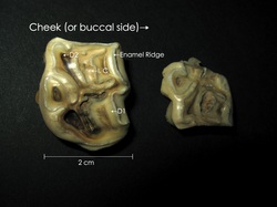
dentin (refer to magnified image of Morning Star's photo). The sharp edges on the
enamel ridges are a very effective
grinding tool for the mastication (chewing) process in horses, which
involves a circular, or elliptical motion (PS Ungar, Ch. 1, Tooth
Structure and Form, Mammal Teeth: Origin, Evolution and Diversity, 2010).
_Occlusal surface of equine permanent premolar. Photograph of 206 occlusal surface. C, cement (light brown); D1, primary dentin
(white/yellowish); D2, secondary dentin overlying pulp horn (dark brown); *, enamel
(visible as a winding ridge); I, infundibulum (infundibular wall enclosing cement core). There are two infundibula in each upper cheek tooth and six pulp horns for 206. The pulp horn is the upper part of the pulp cavity that extends towards the occlusal surface. No information is given as to the age, sex or breed of this horse. Compare to Morning Star's deciduous 206. Caption modified from Lundström et al. (2007), Acta Veterinaria Scandinavica.
_The slope of the occlusal (chewing) surface on the cheek teeth is usually attributed to the anisognathic (Gk, anisos, unequal + gnathos, jaw) nature of the horse's mouth, i.e., the distance between teeth is greater in the upper jaw than in the lower jaw. This difference is thought to produce the wear pattern shown for the premolar annotated in the above image. The opposing premolar in the upper jaw is taller on the buccal side. It has been suggested that the slope is not due entirely to wear (GJ Baker, Dental Physiology, in Equine Dentistry, GJ Baker and J Easley, 2nd Ed., 2005). In this case, the occlusal surface of the tooth is slightly inclined on emergence and develops the greater slope over time due to the grinding action of the cheek teeth. The average slope of the occlusal surface, which is about 10-15° in adult horses, is slightly higher in foals and yearlings, consistent with our measurement in Morning Star.
A recent study of occlusal angles in horse skulls, using malleable wires as imprints rather than a visual assessment, found that this angle is not constant but increases from the rostral (anterior) to the caudal (posterior) position for the mandibular teeth. The opposite trend was found for the maxillary teeth, i.e., smaller angles were observed for the more caudally positioned teeth. In addition, the variation in angle was greater for the lower jaw than for the upper jaw (Brown et al. 2008).
If this relationship is borne out by other studies, the Triadan position of the tooth within the lower and upper arcades (rows of jaw teeth) would have to be considered in dental procedures where the occlusal surface of a cheek tooth must be restored to its original slope.
In the ideal case, horses chew in an elliptical motion producing a uniform wear pattern on the occlusal surfaces of the cheek teeth. When the mediolateral (from middle to side) motion is restricted, however, sharp enamel points can form on the outside edges of the maxillary teeth and the inside edges of the mandibular teeth. Sharp points are usually more common in horses that are fed concentrates and grain, or forage from hanging feeders and mangers, processes that involve a more "up-and-down" chewing motion.
Morning Star is an interesting case because she has never been fed grain or concentrate. Her diet consists totally of hay and grass with a small portion (1/2 quart) of non-molasses beet pulp mixed with a powdered vitamin/trace mineral supplement (McCauley's V-MAX 0.5-oz daily). She has 24/7 access to the outside from her stable, and is always fed off the ground with her head in the grazing position. All our horses are on pasture about 8-10 hours per day, weather permitting.
A recent study published in the journal Animal compares dentition in stabled versus free-grazing Thoroughbreds. As expected, stabled horses had more dental abnormalities. One unexpected result was the common occurrence of sharp enamel points in BOTH free-grazing horses and those that were stabled and fed a simulated grain diet. Based on this observation, the authors pose a rather interesting question: "The results of our study may also suggest that further study is required to decide whether routine removal of sharp edges is actually a necessity and that this process is not removing an anatomical feature that is required by the horse." The implication is that sharp points are not abnormal but a natural feature of the horse's mouth. One can argue that the main reason sharp points are removed is because of the bit!
Exam 2: December 28, 2011
Incisors
|
_
|
__Photos of the labial and occlusal surfaces of deciduous incisors for 12/28/2011 (left) and 04/21/2011 (right) exams. Deciduous incisors are smaller, whiter and more domal in shape than the permanent teeth. At age 2 yrs, 3 mos (exam 2, left), the central incisors have not separated from the gum to form "caps".
|
_
_Relative to exam 1, the cups for the central incisors are smaller and the enamel ring is less elongated (refer to incisor cross section above). Cups on the
intermediates are more shallow, and the corner incisors are in full
wear with a well-defined enamel ring (403). Photos by GA Lager
The above photos compare the dental surfaces of the deciduous incisors photographed during the 12/28/2011 (left) and 04/21/2011 (right) exams. Morning Star's age at the time of the second exam was 2 years and 3 months. Based on the frontal views, the central incisors have not yet been pushed free of the gum by the emerging permanent teeth. The part of the deciduous tooth that is not resorbed (dissolved and assimilated) remains attached to the permanent tooth during eruption and forms a "cap". These "caps" are normally "shed" as the permanent tooth continues to develop. As noted above, permanent incisor 101 erupts on average at about 2.5 years.
The intervening time between exams (~8 months) has produced some additional wear of the occlusal surface. In particular, the cups for the intermediates are shallower and the corner incisors are in full wear with the appearance of an enamel ring. The cups for the lower central incisors are very shallow and have almost disappeared. The enamel ring is oval-shaped but less elongated as the wear extends deeper into the funnel-shaped infundibulum.
The intervening time between exams (~8 months) has produced some additional wear of the occlusal surface. In particular, the cups for the intermediates are shallower and the corner incisors are in full wear with the appearance of an enamel ring. The cups for the lower central incisors are very shallow and have almost disappeared. The enamel ring is oval-shaped but less elongated as the wear extends deeper into the funnel-shaped infundibulum.
Cheek Teeth
No corrections (i.e., rasping) were made to the cheek teeth. The major change since the last exam was the eruption of the second permanent molar (210), which emerges on average at about 2 years of age. Unfortunately, the oblique view angle of the camera makes it difficult to clearly see the permanent molars in the photo. In the diagram below, photos of the maxillary arcades (2nd quadrant) from the last two exams are compared to a perpendicular view of the same arcade cropped from a photo of the palate of an immature horse (photo taken postmortem, Uwe Gille, 2005, Wikimedia Commons, http://creativecommons.org/licenses/by-sa/3.0/). Using this type of "correlation diagram", the second molar (210) can be more readily identified in our photo.
Correlation between photos of the maxillary arcades (2nd quadrant) in the first (left) and second (far right) exams and the same arcade in an "immature horse" (center). The permanent molars in our photos are difficult to see because of the view angle. Left and far right photos by GA Lager
_No information is
given as to the exact age of the "immature horse" but this horse is probably about 4-5 years of age based on the following evidence: 1. the last permanent molar (211) has
erupted, and 2. the central incisors (101 and 201) and one of the intermediate incisors (102) are wide transversely (side to side) with deep cups, indicating that they are permanent teeth (refer to original
uncropped image).
If this age estimate is correct, all permanent cheek teeth have probably erupted (refer to average eruption times in AAEP table).
Note the anatomical differences between the occlusal surfaces of the cheek teeth in the "immature horse" and those in Morning Star. The more resistant enamel ridges in Morning Star's cheek teeth, which are planar with sharp edges, surround deep depressions of softer dentin. In contrast, the ridges on the occlusal surface of the permanent teeth in the "immature horse" are more rounded and some of the depressions are smaller in cross-sectional area and not as deep (e.g., those located in the central part of premolar 206). The lesser degree of wear in the "immature horse" is probably related to a combination of factors, including age, diet and the wear resistance of permanent versus deciduous teeth.
X-ray Radiology
The X-ray absorption in materials is a function of chemical composition, density and thickness. More dense, thicker materials consisting of elements with higher atomic numbers (e.g., bone and enamel), will absorb more X-rays and show a greater degree of whiteness in radiographic images. These materials are described as radio-opaque because less x-rays reach the film, or electronic detector. More radiolucent substances (e.g., fat, tissue and gas cavities) appear darker in radiographs because the absorption is less and more of the X-ray radiation is transmitted. Medical images represented in terms of two-dimensional radiographs are complicated by the fact that there can be a superposition of objects in different planes. In this case, the degree of absorption is summed over several objects.
You can read more about X-ray radiology in the book Veterinary Diagnostic Radiology, by D Thrall, 4th Ed., 2002. It has been scanned and is available in pdf format on the Web. It's a rather large file (116 MB) so if you don't have broadband you are in for a long wait. Chapter 5, Introduction to Radiographic Interpretation, is probably the most useful to horse owners. This earlier edition does not discuss digital radiography, which has replaced conventional film methods in many equine practices. A good online resource based on digital X-ray (computed rather than direct digital radiography) is The Handbook of Equine Radiography published by Elsevier.
We recorded several digital images of Morning Star's mouth to complement our photos. As noted above, this is a very important time in her development because she will shed some of her deciduous teeth within the next several months. To interpret these radiographs, you will need to learn a few anatomical locational terms which describe the positioning of the tube relative to the skull and detector plate. Refer to Fig. 5-17 in Thrall, or to this Wikipedia page.
The frontal view of the skull below shows the orientation of the X-ray beam and detector plate for an oblique radiograph. The description of this orientation (dorsal lateral-ventral axial) sounds very complicated, doesn't it? Actually, all you really need to know to interpret this radiograph is that the X-ray beam passes from the upper right side (dorsal lateral) to the bottom (ventral) of the head at an angle of about 30° to the medial plane (red line in drawing that divides left and right sides of the head). The detector plate (digital radiography), or film cassette, is placed on the opposite side of the head relative to the X-ray tube at 90° to the beam. The view of the radiograph is from the position of the tube. The oblique orientation is useful in separating teeth on opposite sides of the mandible and maxilla and avoiding summation effects due to the superposition of teeth.
_Positioning of the X-ray tube and detector plate in an oblique view of the skull. View is from the front of the horse. Cheek teeth are colored in red. The 3rd and 4th quadrants of the mouth are labeled. Drawing modified from JP Morgan, Techniques of Veterinary Radiology, 5th Ed., 1993.
_The digital radiograph
shows several of the mandibular incisors and the first two premolars in
the 4th quadrant of the mouth (refer to figure above on the Triadan
numbering system). However, summation effects are not entirely
eliminated. For example, the incisors circled in red represent
superposition of teeth in the 3rd and 4th quadrants.
_This radiograph is not very informative in terms of the spatial relationship between the deciduous and permanent incisors.
The University of Illinois College of Veterinary Medicine has compiled interactive medical images of the equine skull of a 2-year old Thoroughbred filly. Click on the tabs at the top of the page for color renditions of the different types of teeth. The first radiograph in this series shows the development of the permanent incisors on the lingual (tongue) side of the deciduous teeth. At age 2 years and 3 months, Morning Star's permanent incisors are probably at a similar stage of development.
The University of Illinois College of Veterinary Medicine has compiled interactive medical images of the equine skull of a 2-year old Thoroughbred filly. Click on the tabs at the top of the page for color renditions of the different types of teeth. The first radiograph in this series shows the development of the permanent incisors on the lingual (tongue) side of the deciduous teeth. At age 2 years and 3 months, Morning Star's permanent incisors are probably at a similar stage of development.
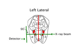
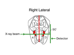
Our next two radiographs are lateral views from the left and right sides of the skull. In this type of radiograph, the X-ray tube is positioned so that the beam is perpendicular to both the medial plane and the detector on the opposite side of the skull. Lateral radiographs are generally not as helpful because of the superposition of teeth on opposites sides of the jaws (see diagrams below).
_Positioning of the X-ray tube and detector plate in lateral views of the skull. View is from the front of the horse. Cheek teeth are colored in red. The 3rd and 4th quadrants of the mouth are labeled. Drawings modified from JP Morgan, Techniques of Veterinary Radiology, 5th Ed., 1993.
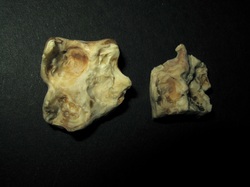
In the left lateral below, the area outlined in red shows the deciduous "caps" overlying permanent premolars 306 and 307. The "caps", remnants of the deciduous teeth, are more radiolucent than the permanent teeth because they are less dense and, therefore, absorb less X-rays. Compare the premolar deciduous "caps" in this radiograph to those in the skull lateral of the young Thoroughbred filly.
A recent study of 207 Thoroughbred racehorses by Ramzan et al. (2009) found that eruption times of permanent premolars based on age at "cap" removal differed significantly from those published in the equine literature. Mean ages at "cap" removal were 35.1, 37.7 and 45.1 months for premolars 206, 207 and 208, respectively. The corresponding eruption times from the AAEP table are 30, 36 and 48 months. The results also showed that the age at "cap" removal increased for more caudal teeth, teeth in the upper jaw and for mares.
A recent study of 207 Thoroughbred racehorses by Ramzan et al. (2009) found that eruption times of permanent premolars based on age at "cap" removal differed significantly from those published in the equine literature. Mean ages at "cap" removal were 35.1, 37.7 and 45.1 months for premolars 206, 207 and 208, respectively. The corresponding eruption times from the AAEP table are 30, 36 and 48 months. The results also showed that the age at "cap" removal increased for more caudal teeth, teeth in the upper jaw and for mares.
Views of the top and bottom of two deciduous "caps" from cheek teeth of a Thoroughbred racehorse, Churchill Downs, Louisville, Kentucky. "Caps" provided by Dr. Atteyat Hadizadeh. A thin shell attached to the occlusal surface of the emerging permanent tooth is all that remains of the deciduous tooth after resorption. Labels: I, infundibulum; C, cement (yellowish brown); D1, primary dentin (white/yellowish); D2, secondary dentin overlying pulp horn (dark brown). The larger "cap" is approximately 3 x 2 x 0.8 cm. Photos by GA Lager
_The rounded,
radiolucent area circled in red at the apex of 307 represents an
eruption cyst, or teething bump, located at the bottom of the mandible
(SZ Barakzai, Ch. 13, Dental Imaging, in Equine Dentistry,
J Easley, PM Dixon and J Schumacher, 3rd Ed., 2011). The corresponding
teething bump on the opposite side of the mandible (407) is shown in the
oblique view. Eruption cysts
form as the tooth bud develops into the permanent cheek tooth. As
mentioned in the section on Exam 1, the first teething bump, which
disappeared about one month prior to this exam, was probably associated
with 306.
The two permanent molars (409 and 410) and the deciduous premolar (408) in the mandible are labeled in the right lateral. Both molars are erupted and in wear. There is a "cap" present on 408, which is the last permanent cheek tooth to erupt (~ 4 years). The angulation of the cheek teeth (red lines) creates a closely packed row, or arcade, of teeth, as shown in the photos above. Compare to Fig. 6.1, JL Carmalt, Ch. 6, Dental Physiology, in Equine Dentistry, J Easley, PM Dixon and J Schumacher, 3rd Ed., 2011.
Exam 3: May 20, 2012
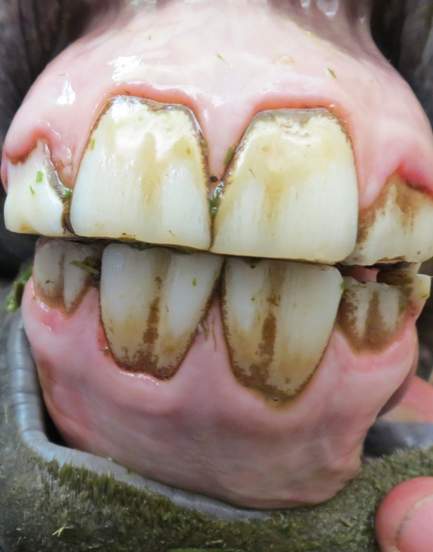
The objectives of Exam 3 were to monitor the development of the permanent teeth and examine wear patterns on the occlusal surfaces of the cheek teeth. As shown in the photo below, all four of the permanent central incisors (101) have emerged at 2 years and 8 months. The timing of our exam was such that we were able to observe the shedding of the deciduous "cap" on incisor 301. The deciduous "caps" on the second premolar, or first cheek tooth, have not been shed (average eruption time 2.5 years).
No adjustments were made to the cheek teeth. Unusual wear patterns, or sharp enamel points, were not observed.
No adjustments were made to the cheek teeth. Unusual wear patterns, or sharp enamel points, were not observed.
Eruption of permanent central incisors and shedding of 301 "cap". The permanent teeth are larger with vertical grooves. Compare to images of deciduous incisors in Exam 2. The permanent incisors are not in wear at this age. Photo by GA Lager
View of the cheek teeth in the 2nd and part of the 3rd quadrant. The deciduous "caps" on premolars 206 and 306 (first tooth in arcade) have not been shed. The small white premolar (205) is the wolf tooth. Photo by GA Lager
Exam 4: October 4, 2012
Incisors
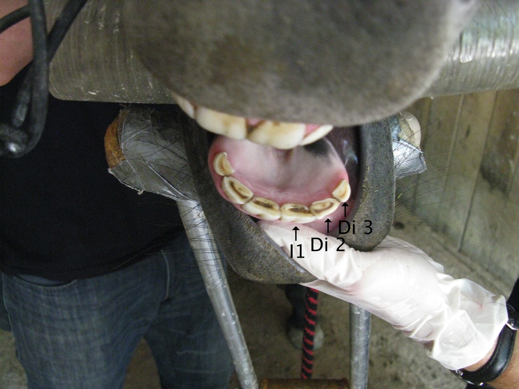
The convention for labeling deciduous teeth recommended by the AAEP has been adopted in this exam (Guide for Determining the Age of the Horse, American Association of Equine Practitioners, 2010). It simplifies labeling of images when arcades of incisors and cheek teeth are composed of both deciduous and permanent teeth (I = permanent incisor; P = permanent premolar; Di or D = deciduous tooth; p = deciduous premolar, e.g., P2 = permanent second premolar; Dp2 = deciduous second premolar).
The two permanent central incisors (I1) noted in the previous exam are now in full wear at age 3 years and 1 month. The intermediates and corners are deciduous (Di 2 and Di 3).
The two central incisors are permanent and in full wear. The intermediate and corners are deciduous (Di). Photo by GA Lager
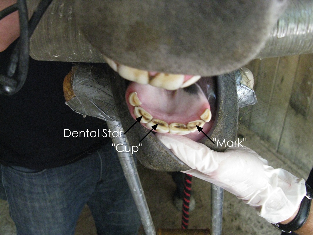
The permanent teeth have well developed "cups" and are larger than the deciduous teeth. The "cups" have disappeared in Di 2 and Di 3, which are shed on average at about 3.5 and 4.5 years, respectively. The enamel ring in the deciduous teeth now surrounds cement in the lower part of the infundibulum and is referred to as the "mark". Although it is difficult to determine from this photo, the small round feature in the middle of the cement core (e.g., Di 2) is probably a remnant of the "cup".
The dental star is present in both Di 2 and Di 3. This feature appears around 9 years of age in the permanent central incisors based on the generalized diagram shown in Exam 1 (Anatomy of the equine permanent incisor). In reality, the appearance of the dental star with age in permanent incisors is quite variable and depends on the anatomy of the tooth, e.g., the depth of dentin overlying the pulp cavity (see, for example, S Muylle, Ch. 7, Aging, in Equine Dentistry, J Easley, PM Dixon and J Schumacher, 3rd Ed., 2011).
The dental star is present in both Di 2 and Di 3. This feature appears around 9 years of age in the permanent central incisors based on the generalized diagram shown in Exam 1 (Anatomy of the equine permanent incisor). In reality, the appearance of the dental star with age in permanent incisors is quite variable and depends on the anatomy of the tooth, e.g., the depth of dentin overlying the pulp cavity (see, for example, S Muylle, Ch. 7, Aging, in Equine Dentistry, J Easley, PM Dixon and J Schumacher, 3rd Ed., 2011).
The permanent central incisors have deep "cups". The "cups" are no longer present in the deciduous intermediate and corner incisors. The occlusal surface in Di 2 and Di 3 is characterized by the presence of the "mark", in which the enamel ring surrounds cement in the lower part of the infundibulum. The small round feature on the occlusal surface of Di 2 (tip of arrowhead) is probably a remnant of the "cup". The dental star appears on the labial side of the "mark". Photo by GA Lager
The raised appearance of the soft tissue above Di 2 (102) indicates that this incisor will soon separate from the gum as the permanent tooth continues to erupt.
The permanent central incisors are larger than the deciduous intermediates and corners, which is illustrated in this image of the maxillary incisors. Di 2 (102) is about to separate from the gum and will be shed within the next few months (average eruption time 3.5 years). Note the raised area just above the gum line. Photo by GA Lager
Our photos are similar to those shown on page 17 of the AAEP Guide for Determining the Age of the Horse.
Cheek Teeth
The second premolar (P2) has erupted since the last exam. Five of the six pulp horns and the two infundibula can be clearly distinguished in the image below. Compare its occlusal surface with those in previous exams. Note the the more rounded (rather than planar) shape of the enamel ridges and the poorly developed depressions in the dentin above the pulp cavities, consistent with the time of eruption and the degree of wear.
If you look closely at this image you can also see the eruption of P3 (107) below deciduous "cap" Dp3. The margin between P3 and Dp3 is indicated by the red arrow.
If you look closely at this image you can also see the eruption of P3 (107) below deciduous "cap" Dp3. The margin between P3 and Dp3 is indicated by the red arrow.
A portion of the maxillary arcade in 1st quadrant showing the wolf tooth (P1), permanent second premolar (P2), permanent third premolar (P3) and its deciduous "cap" (Dp3). The margin between P3 and Dp3 is indicated by the red arrow. Also note the resorption feature on Dp3 at the end and to the left of this arrow opposite the arrowhead. Photo by GA Lager
This second image shows Dp3 in the second quadrant. This "cap" was loose on examination and could have been removed. In this project, however, our goal was to monitor rather than extract deciduous "caps".
The fourth deciduous premolar Dp4 is also visible.
P1 (205 or wolf tooth) is no longer present and was shed during eruption of the permanent second premolar (P2 or 206). In some cases, when the wolf tooth is located close to the second premolar, P2 eruption can cause resorption of the P1 root (J Easley, Ch. 17, Corrective Dental Procedures, in Equine Dentistry, J Easley, PM Dixon and J Schumacher, 3rd Ed., 2011). Note that 205 was present in Exam 3 before the deciduous "cap" of 206 was shed.
The P3 (207) deciduous cap (Dp3) was loose on examination but was not removed. The opposing premolar in the mandible (307) is permanent and already in wear. Photo by GA Lager
The view below shows the maxillary arcade of cheek teeth. At this age (3 years and 1 month), all permanent molars and premolars have erupted in the upper and lower jaws with the exception of fourth premolar (P4), which is usually the last to erupt (~4 years). The three molars at the back of mouth are best viewed at highest magnification (two mouse clicks). As mentioned, one of the wolf teeth (205) was shed during eruption of P2.
All permanent cheek have erupted with the exception of P4, the fourth premolar. The wolf tooth (205) was shed during eruption of P2. Magnify this image for a good look at the three molars (209-211). Photo by GA Lager
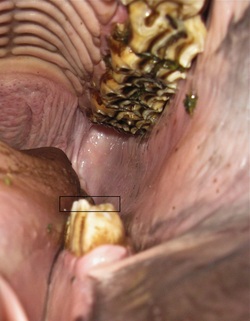
A slight medial rise, i.e., toward the middle of the mouth, was observed on the occlusal surfaces of 307 and Dp4 (308), as shown below in the black rectangle. Based on personal observation (SH Christie), enamel ridges are softer on the occlusal surface of newly erupted molars and become harder with age. Because 307 erupts about 6 months before 207 (see X-ray radiograph of 307, Exam 2, December 28, 2011), uneven wear might be expected until opposing permanent teeth are in occlusion (in contact).
The medial rise was not corrected by rasping because Morning Star will be trained without a bit using the Scawbrig bitless bridle (see Bitless Bridles Page). The same reasoning applies to the wolf teeth, which were not extracted.
The medial rise was not corrected by rasping because Morning Star will be trained without a bit using the Scawbrig bitless bridle (see Bitless Bridles Page). The same reasoning applies to the wolf teeth, which were not extracted.
Uneven wear on 307 and Dp4 (black rectangle). Not corrected but will be monitored during training with bitless bridle. Wear pattern for 307 might be affected by eruption time, i.e., the opposing permanent tooth in maxilla (207) erupted about 6 months after 307. Photo by GA Lager
Equine Dental Record 10.04.2012
Because some of you have expressed interest in seeing an actual dental exam chart, we have published the 10.04.2012 exam results. The intent of the exam is to identify developmental abnormalities, which might require corrective dental procedures.
The only abnormality observed was the slight medial rise on the occlusal surfaces of 307 and 308 (Dp4). This was not corrected but will be monitored for the reasons cited above.
The loose “caps” on 107 and 207 are part of the developmental process and are normally not removed unless the horse is in training with a bit, or the “caps” are not shed when the permanent tooth erupts (retained “cap”). The “cap” can also become painful if impacted with food or fractured, necessitating removal.
The shedding of wolf teeth during eruption of the second premolar is not uncommon, judging from the lower percentage of wolf teeth in adult versus younger (~2 years) horses (J Easley, Ch. 17, Corrective Dental Procedures, in Equine Dentistry, 3rd Ed., 2011).
Note that some of the other recommended corrective procedures (e.g., rounding of the rostral (front) corners of the second premolar, etc.) are unnecessary if a bit is not used. Refer to J Easley, Ch. 17, Equine Dentistry for exam recommendations for younger horses.
The exam form shown is available through Bellwether Publishing Inc.
Exam 5: April 24, 2013
This exam focused on the further development of incisors and eruption of last cheek tooth, the fourth premolar. Morning Star's age at the time of this exam was 3 years and 7 months.
Incisors
Development of incisors since Exam 4. Intermediate incisors (302 and 402) in mandible have shed caps; those in maxilla (102 and 202) are about to be shed (see separation at gum line). Central incisors (301 and 401) show more wear but "cups" are still deep. Large, well-defined "cups" in intermediate incisors (302 and 402), which are not in wear. Note larger size and vertical grooves and ridges of permanent teeth relative to deciduous corners. The photo on the left was taken during the radiographic exam on 05.02.2013. Photos by GA Lager
Cheek Teeth
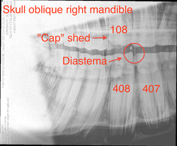
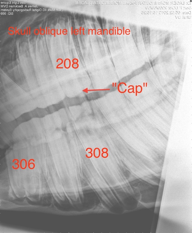
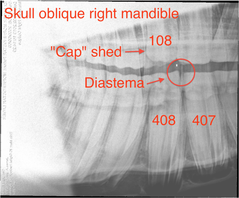
Eruption of fourth premolar. Oblique X-ray radiographs of right and left sides of skull showing 408 diastema (space, or gap, between two teeth) and development of fourth premolars in maxillary and mandibular arcades. Diastema is within "normal limits" and is developmental (e.g., teeth angulation and spacing) rather than pathological (caused by disease) in nature. Although this tooth will be monitored on a regular basis, no future problems (e.g., periodontal disease from food packing) are expected. For more information on diastemata in cheek teeth refer to AAEP focus paper by Dixon (2006). The deciduous "cap" on the fourth premolar (108) has recently been shed (arrow). Premolar 408 is not in occlusion, which might account, in part, for overgrowth at rostral end of tooth adjacent to diastema. Both the third and fourth premolars in the mandible erupt about six months before opposing teeth in the maxilla. Digital radiographs, Dr. James Beckman, Gas Light Equine
Image of second and third quadrants showing former site of wolf tooth (205) and location of medial rise observed for 307 in Exam 4. The soft tissue protrusion from gum after shedding of 205 was not observed in this exam (see above). The medial rise appears to be less steep after eruption of 207 and occlusion of opposing premolars. Photo by GA Lager
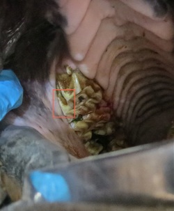
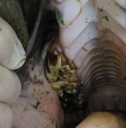
Images showing the effect of rasping with a power tool on sharp points and edges on second premolar 106. The remaining wolf tooth (105) is also shown. The origin of the small puncture wound on the lingual side of 106/107 is unknown and was not related to the dental exam. Compare buccal side of 106 within the red rectangle before (left) and after rasping. Note how points and edges have been rounded in photo on the right, taken during radiographic exam on 05.02.2013. This is a conservative "float" designed to minimize potential problems during training without jeopardizing the horse's ability to chew forage efficiently. Photos by GA Lager
Exam 6: November 20, 2013
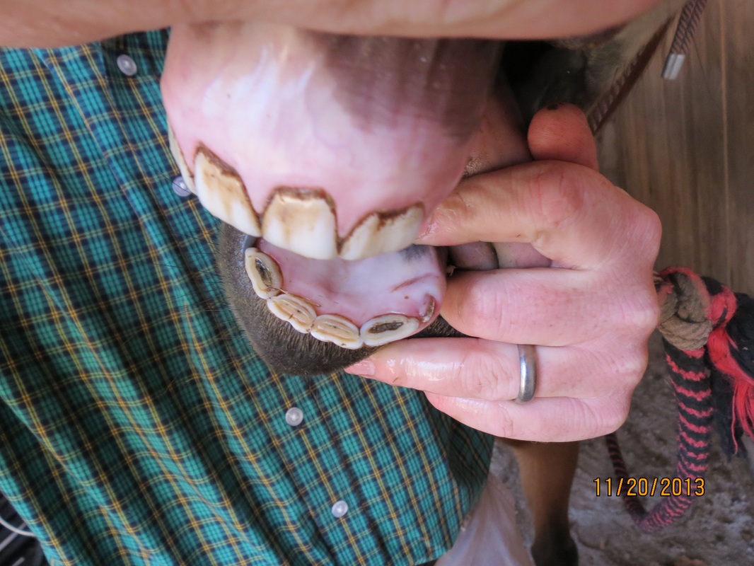
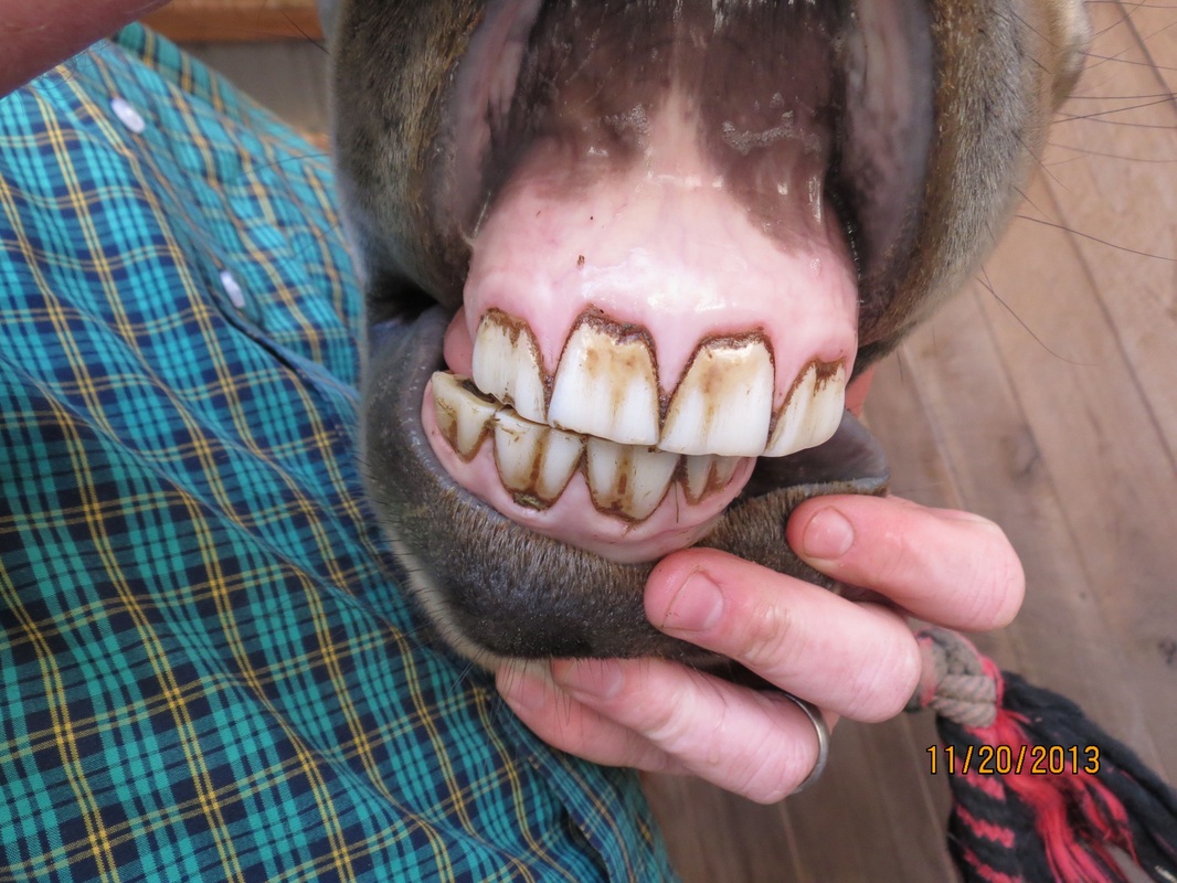
Shortly after the radiographic exam on 05.02.2013, Morning Star fractured and lost both deciduous lower corner incisors (303 and 403). The premature loss of 403 was by chance observed directly. A vertical fracture developed along the midline in 403. One half of the tooth was lost, followed shortly by the second half. All that remains above gum line are small fragments. The cause of the fracture is unknown but might be related to her habit of occasionally grasping the metal rails of the corral panels and sliding her teeth along the length of the rails (a bad habit she picked up from her mom). The upper deciduous corners are still intact.
One objective of the current exam was to determine the effect of loss of these incisors on development of the permanent teeth. At her present age (4 years 2 months), Morning Star is within the time window for eruption of the permanent corner incisors (4-5 years).
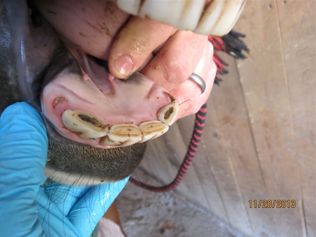
The second objective was to monitor the development of the fourth premolar, the last permanent cheek tooth to erupt. In the last exam, visual examination and radiographs revealed a developmental diastema between 407 and 408. The deciduous “cap” had just been shed on the opposing maxillary premolar 108 and 108/408 were not in occlusion.
Incisor photographs
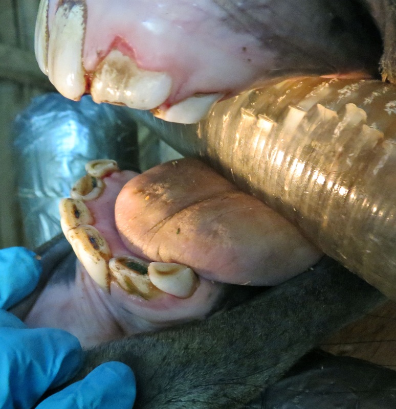
Normal (left) and oblique views of the lower incisors showing the remaining fragments of the deciduous corners. Relative to the last exam the permanent intermediate incisors are in full wear and have well-developed, deep "cups". The occlusal surfaces of the central incisors are essentially smooth with shallow "cups". The dental star has not yet appeared in the central incisors, which have been in wear for almost two years. Photos by GA Lager
Frontal view of the four permanent incisors. The upper deciduous corner incisors are still intact (not visible). Photo by GA Lager
X-ray Radiography
Incisors
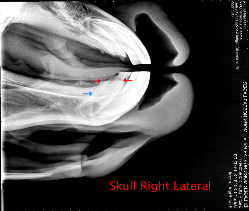
Right lateral showing the projection of 303 and 403 on the radiographic plane. The two permanent corner incisors are slightly offset in projection, as indicated by the parallel lines on lingual side (blue arrow). The two opposing red arrows define the line of intersection between the deciduous crown below gum line and the developing permanent tooth. The deciduous teeth are radiolucent and not readily visible, in contrast to the bright image of the more dense, radio-opaque permanent teeth. Based on the radiographic evidence, the premature loss of the deciduous corner incisors will have minimal effect on the eruption of the permanent counterparts. Digital radiographs, Dr. James Beckman, Gas Light Equine
Horse owners rarely have the opportunity to see the occlusal surfaces of the maxillary (upper) incisors during a dental exam. In the intraoral dorsoventral projection, the detector plate, or cassette, is placed in the mouth perpendicular to X-ray beam, which is focused at a point above the incisor roots. The four permanent incisors produce a bright X-ray image relative to the deciduous corner incisors. The enamel rings and central "cups", which are composed of materials with different densities, show good contrast in X-ray images (refer to drawing of incisor anatomy in Exam 1). Compare to photo of mandibular incisors above. Digital radiographs, Dr. James Beckman, Gas Light Equine
Development of permanent fourth premolar
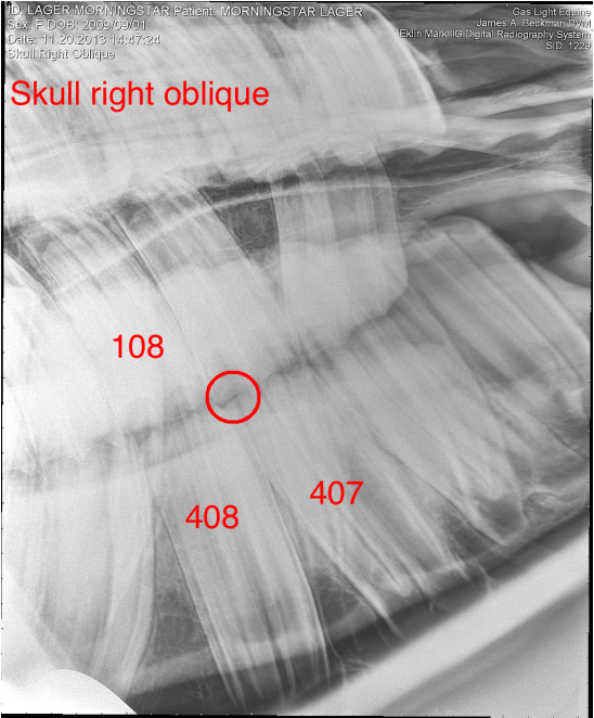
Skull radiograph from Exam 5 on 05.02.2013 showing relationship between permanent premolars 108 and 408. The deciduous cap on 108 had been shed recently and the tooth was not in wear. The diastema between 408 and 407 is circled in red. Digital radiographs, Dr. James Beckman, Gas Light Equine
In present exam, seven months later, premolars 108 and 408 are in occlusion and the diastema (red circle) can no longer be identified in the radiograph. Digital radiographs, Dr. James Beckman, Gas Light Equine
June 2014 Update to Exam 6
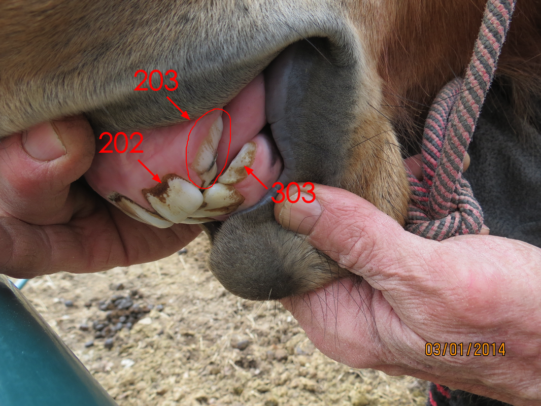
The development of the permanent lower and upper corner incisors have been monitored since the last exam. As shown in the images below, the lower corners erupted normally, as predicted from the X-ray radiographs recorded about six months ago. However, an extra tooth was observed in the upper incisor arcade in both the 103 and 203 positions.
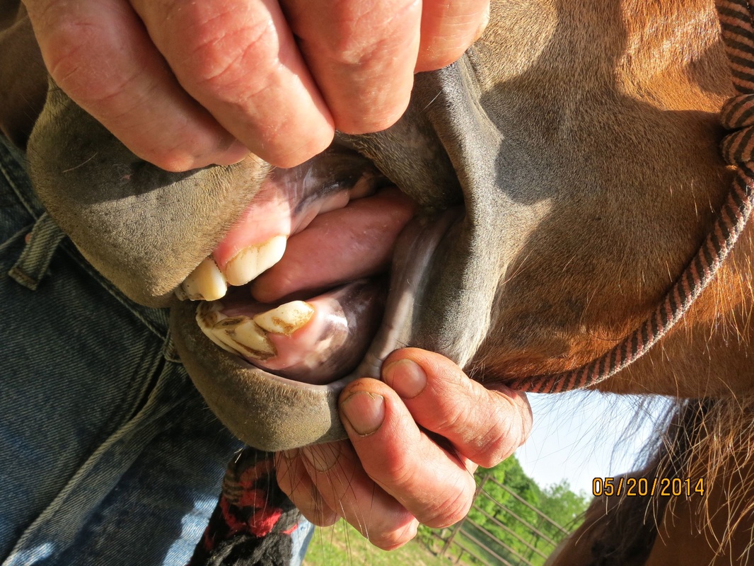
Lateral views of the second and third quadrants of Morning Star's incisor arcade taken in March (left) and May (right) of this year. Note normal development of the permanent lower corner incisor (303) and additional smaller tooth located on the lingual (palatal) side in upper incisor arcade (203). The smaller tooth is most likely a retained deciduous incisor as opposed to a supernumerary tooth (additional permanent tooth). The size and crown shape of the latter are usually the same as those of the normal permanent teeth. In May photo smaller tooth is present but hidden from view. This preliminary diagnosis will be further investigated in upcoming dental exam. Refer to articles by Dixon (2005) on dental disorders and Dixon et al. (2005) on supernumerary teeth for more information.
Lateral view showing the development of the corner incisor (203) after about 4 months since the last photo. Breadths of the corner incisors in the upper jaw (103 and 203) are
anomalously wide. Occlusal surfaces of both corners overlap those of the lower intermediate incisors. Smaller tooth on the palatal side could not found on palpation; however, conclusive evidence for the absence of this tooth requires a thorough dental exam, preferably with a oral camera, or dental mirror.
anomalously wide. Occlusal surfaces of both corners overlap those of the lower intermediate incisors. Smaller tooth on the palatal side could not found on palpation; however, conclusive evidence for the absence of this tooth requires a thorough dental exam, preferably with a oral camera, or dental mirror.
Copyright George Lager, 2011-2024 All Rights Reserved (®)


